Legg–Calvé–Perthes disease (LCPD) is a childhood hip disorder initiated by a disruption of blood flow to the head of the femur. Due to the lack of blood flow, the bone dies and stops growing. Over time, healing occurs by new blood vessels infiltrating the dead bone and removing the necrotic bone which leads to a loss of bone mass and a weakening of the femoral head.

Ainhum, also known as dactylolysis spontanea, is a painful constriction of the base of the fifth toe frequently followed by bilateral spontaneous autoamputation a few years later.
Dysbaric osteonecrosis or DON is a form of avascular necrosis where there is death of a portion of the bone that is thought to be caused by nitrogen (N2) embolism (blockage of the blood vessels by a bubble of nitrogen coming out of solution) in divers. Although the definitive pathologic process is poorly understood, there are several hypotheses:

Avascular necrosis (AVN), also called osteonecrosis or bone infarction, is death of bone tissue due to interruption of the blood supply. Early on, there may be no symptoms. Gradually joint pain may develop, which may limit the person's ability to move. Complications may include collapse of the bone or nearby joint surface.

Kienböck's disease is a disorder of the wrist. It is named for Dr. Robert Kienböck, a radiologist in Vienna, Austria who described osteomalacia of the lunate in 1910.

In vertebrate anatomy, the hip, or coxa in medical terminology, refers to either an anatomical region or a joint on the outer (lateral) side of the pelvis.

Slipped capital femoral epiphysis is a medical term referring to a fracture through the growth plate (physis), which results in slippage of the overlying end of the femur (metaphysis).
Osteochondrosis is a family of orthopedic diseases of the joint that occur in children, adolescents and rapidly growing animals, particularly pigs, horses, dogs, and broiler chickens. They are characterized by interruption of the blood supply of a bone, in particular to the epiphysis, followed by localized bony necrosis, and later, regrowth of the bone. This disorder is defined as a focal disturbance of endochondral ossification and is regarded as having a multifactorial cause, so no one thing accounts for all aspects of this disease.

Osteochondritis dissecans is a joint disorder primarily of the subchondral bone in which cracks form in the articular cartilage and the underlying subchondral bone. OCD usually causes pain during and after sports. In later stages of the disorder there will be swelling of the affected joint that catches and locks during movement. Physical examination in the early stages does only show pain as symptom, in later stages there could be an effusion, tenderness, and a crackling sound with joint movement.
Hypertrophic Osteodystrophy (HOD) is a bone disease that occurs most often in fast-growing large and giant breed dogs; however, it also affects medium breed animals like the Australian Shepherd. The disorder is sometimes referred to as metaphyseal osteopathy, and typically first presents between the ages of 2 and 7 months. HOD is characterized by decreased blood flow to the metaphysis leading to a failure of ossification and necrosis and inflammation of cancellous bone. The disease is usually bilateral in the limb bones, especially the distal radius, ulna, and tibia.

The obturator artery is a branch of the internal iliac artery that passes antero-inferiorly on the lateral wall of the pelvis, to the upper part of the obturator foramen, and, escaping from the pelvic cavity through the obturator canal, it divides into an anterior branch and a posterior branch.

Transient synovitis of hip is a self-limiting condition in which there is an inflammation of the inner lining of the capsule of the hip joint. The term irritable hip refers to the syndrome of acute hip pain, joint stiffness, limp or non-weightbearing, indicative of an underlying condition such as transient synovitis or orthopedic infections. In everyday clinical practice however, irritable hip is commonly used as a synonym for transient synovitis. It should not be confused with sciatica, a condition describing hip and lower back pain much more common to adults than transient synovitis but with similar signs and symptoms.

Osteonecrosis of the jaw (ONJ) is a severe bone disease (osteonecrosis) that affects the jaws. Various forms of ONJ have been described since 1861, and a number of causes have been suggested in the literature.

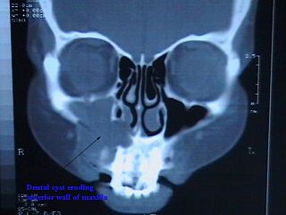
Commonly known as a dental cyst, the periapical cyst is the most common odontogenic cyst. It may develop rapidly from a periapical granuloma, as a consequence of untreated chronic periapical periodontitis.

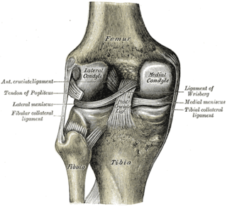
Spontaneous osteonecrosis of the knee is the result of vascular arterial insufficiency to the medial femoral condyle of the knee resulting in necrosis and destruction of bone. It is often unilateral and can be associated with a meniscal tear.

Chandler's disease, also known as idiopathic avascular osteonecrosis of the femoral head, is a rare condition in which the bone cells in the head of the femur (FH) die due to lack of blood. This disease is caused when blood flow is reduced to the part of a bone near a joint. It is specifically unique because the femoral head is for some reason the only affected part of the body and rarely travels down to the main part of the femur. In 1948, F. A. Chandler did a multi-case review and first released his interpretations as Coronary Disease of the Hip. This term is now considered incorrect as it improperly describes the actual disease.

Pain in the hip is the experience of pain in the muscles or joints in the hip/ pelvic region, a condition commonly arising from any of a number of factors. Sometimes it is closely associated with lower back pain.
An occult fracture is a fracture that is not readily visible, generally in regard to projectional radiography ("X-ray"). Radiographically, occult and subtle fractures are a diagnostic challenge. They may be divided into 1) high energy trauma fracture, 2) fatigue fracture from cyclical and sustained mechanical stress, and 3) insufficiency fracture occurring in weakened bone. Independently of the cause, the initial radiographic examination can be negative either because the findings seem normal or are too subtle. Advanced imaging tools such as computed tomography, magnetic resonance imaging (MRI), and scintigraphy are highly valuable in the early detection of these fractures.

A Phemister graft is a type of bone graft which uses bone tissue harvested from the patient to treat slow-healing, or delayed union bone fractures. Thus, it is a form of autotransplantation. Typically, the tissue used in the graft is cancellous bone harvested from the patient's Iliac crest and laid in strips across the fracture site. The use of the patient's living bone stimulates osteogenesis, the growth of bones.
Dieterich's disease, also known as avascular necrosis of the metacarpal head, is an extremely rare condition characterized by temporary or permanent loss of blood supply to the metacarpal head of the metacarpal bone, resulting in loss of bone tissue. The five metacarpal bones are long bones located between the carpals of the wrist and phalanges of the fingers. Collectively, the metacarpals are referred to as the "metacarpus."


