
Macrocephaly is a condition in which circumference of the human head is abnormally large. It may be pathological or harmless, and can be a familial genetic characteristic. People diagnosed with macrocephaly will receive further medical tests to determine whether the syndrome is accompanied by particular disorders. Those with benign or familial macrocephaly are considered to have megalencephaly.

A cephalohematoma, also spelled cephalohaematoma, is a hemorrhage of blood between the skull and the periosteum at any age, including a newborn baby secondary to rupture of blood vessels crossing the periosteum. Because the swelling is subperiosteal, its boundaries are limited by the individual bones, in contrast to a caput succedaneum.

A periosteal reaction is the formation of new bone in response to injury or other stimuli of the periosteum surrounding the bone. It is most often identified on X-ray films of the bones.

Hypophosphatasia (; also called deficiency of alkaline phosphatase, phosphoethanolaminuria, or Rathbun's syndrome; sometimes abbreviated HPP) is a rare, and sometimes fatal, inherited metabolic bone disease. Clinical symptoms are heterogeneous, ranging from the rapidly fatal, perinatal variant, with profound skeletal hypomineralization, respiratory compromise or vitamin B6 dependent seizures to a milder, progressive osteomalacia later in life. Tissue non-specific alkaline phosphatase (TNSALP) deficiency in osteoblasts and chondrocytes impairs bone mineralization, leading to rickets or osteomalacia. The pathognomonic finding is subnormal serum activity of the TNSALP enzyme, which is caused by one of 388 genetic mutations identified to date, in the gene encoding TNSALP. Genetic inheritance is autosomal recessive for the perinatal and infantile forms but either autosomal recessive or autosomal dominant in the milder forms.
Periostitis, also known as periostalgia, is a medical condition caused by inflammation of the periosteum, a layer of connective tissue that surrounds bone. The condition is generally chronic and is marked by tenderness and swelling of the bone and pain.
Pachydermoperiostosis (PDP) is a rare genetic disorder that affects both bones and skin. Other names are primary hypertrophic osteoarthropathy or Touraine-Solente-Golé syndrome. It is mainly characterized by pachyderma, periostosis and finger clubbing.
A lipid storage disorder is any one of a group of inherited metabolic disorders in which harmful amounts of fats or lipids accumulate in some body cells and tissues. People with these disorders either do not produce enough of one of the enzymes needed to metabolize and break down lipids or, they produce enzymes that do not work properly. Over time, the buildup of fats may cause permanent cellular and tissue damage, particularly in the brain, peripheral nervous system, liver, spleen, and bone marrow.

Laryngomalacia is the most common cause of chronic stridor in infancy, in which the soft, immature cartilage of the upper larynx collapses inward during inhalation, causing obstruction of the airways. It can also be seen in older patients, especially those with neuromuscular conditions resulting in weakness of the muscles of the throat. However, the infantile form is much more common. Laryngomalacia is one of the most common laryngeal congenital disease in infancy and public education about the signs and symptoms of the disease is lacking.
Craniomandibular osteopathy, also known as lion's jaw, is a developmental disease in dogs causing extensive bony changes in the mandible and skull. In this disease, a cyclical resorption of normal bone and replacement by immature bone occurs along the inner and outer surfaces of the affected bones. It usually occurs between the ages of 3 and 8 months. Breeds most commonly affected include the West Highland White Terrier, Scottish Terrier, Cairn Terrier, and Boston Terrier. It is rare in large-breed dogs, but it has been reported. Symptoms include firm swelling of the jaw, drooling, pain, and difficulty eating.
An osteochondrodysplasia, or skeletal dysplasia, is a disorder of the development of bone and cartilage. Osteochondrodysplasias are rare diseases. About 1 in 5,000 babies are born with some type of skeletal dysplasia. Nonetheless, if taken collectively, genetic skeletal dysplasias or osteochondrodysplasias comprise a recognizable group of genetically determined disorders with generalized skeletal affection. These disorders lead to disproportionate short stature and bone abnormalities, particularly in the arms, legs, and spine. Skeletal dysplasia can result in marked functional limitation and even mortality.
Neonatal-onset multisystem inflammatory disease is a rare genetic periodic fever syndrome which causes uncontrolled inflammation in multiple parts of the body starting in the newborn period. Symptoms include skin rashes, severe arthritis, and chronic meningitis leading to neurologic damage. It is one of the cryopyrin-associated periodic syndromes.

The endosteum is a thin vascular membrane of connective tissue that lines the inner surface of the bony tissue that forms the medullary cavity of long bones.

Osteoblastoma is an uncommon osteoid tissue-forming primary neoplasm of the bone.

The Codman triangle is the triangular area of new subperiosteal bone that is created when a lesion, often a tumor, raises the periosteum away from the bone. A Codman triangle is not actually a full triangle. Instead, it is often a pseudotriangle on radiographic findings, with ossification on the original bone and one additional side of the triangle, which forms a two sided triangle with one open side. This two sided appearance is generated due to a tumor that is growing at a rate which is faster than the periosteum can grow or expand, so instead of dimpling, the periosteum tears away and provides ossification on the second edge of the triangle. The advancing tumour displaces the periosteum away from the bone medulla. The displaced and now lateral periosteum attempts to regenerate underlying bone. This describes a periosteal reaction.
Diffuse infantile fibromatosis is a rare condition affecting infants during the first three years of life. This condition is a multicentric infiltration of muscle fibers with fibroblasts resembling those seen in aponeurotic fibromas, presenting as lesions and tumors confined usually to the muscles of the arms, neck, and shoulder area Diffuse infantile fibromatosis is characterized by fast growing benign tumors. This disorder is known to be caused by mutations in germline variants, PDGFRB and NOTCH3, which may be generationally-inherited through autosomal dominant and recessive traits. Although diffuse infantile fibromatosis is classified as benign, it can still lead to life-threatening complications and damage other organs.
A child bone fracture or a pediatric fracture is a medical condition in which a bone of a child is cracked or broken. About 15% of all injuries in children are fracture injuries. Bone fractures in children are different from adult bone fractures because a child's bones are still growing. Also, more consideration needs to be taken when a child fractures a bone since it will affect the child in his or her growth.
Osteomyelitis of the jaws is osteomyelitis which occurs in the bones of the jaws. Historically, osteomyelitis of the jaws was a common complication of odontogenic infection. Before the antibiotic era, it was frequently a fatal condition.
Glomerulocystic kidney disease (GCKD) is a cystic disorder of the kidneys. GCKD involves cystic dilation of Bowman's capsule. It can occur with or without congenital abnormality.
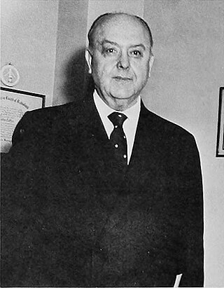
John Patrick Caffey was an American pediatrician and radiologist who is often referred to as one of the founders of pediatric radiology. He was the first to describe shaken baby syndrome, infantile cortical hyperostosis, and Kenny-Caffey syndrome.

Calvarial doughnut lesions-bone fragility syndrome, also known as familial calvarial doughnut lesions, is a rare autosomal dominant genetic disorder characterized by mild to moderate fragility of the bones accompanied with calvarial doughnut lesions.
