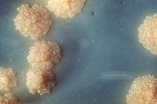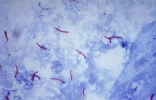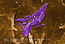
Mycobacterium tuberculosis, also known as Koch's bacillus, is a species of pathogenic bacteria in the family Mycobacteriaceae and the causative agent of tuberculosis. First discovered in 1882 by Robert Koch, M. tuberculosis has an unusual, waxy coating on its cell surface primarily due to the presence of mycolic acid. This coating makes the cells impervious to Gram staining, and as a result, M. tuberculosis can appear weakly Gram-positive. Acid-fast stains such as Ziehl–Neelsen, or fluorescent stains such as auramine are used instead to identify M. tuberculosis with a microscope. The physiology of M. tuberculosis is highly aerobic and requires high levels of oxygen. Primarily a pathogen of the mammalian respiratory system, it infects the lungs. The most frequently used diagnostic methods for tuberculosis are the tuberculin skin test, acid-fast stain, culture, and polymerase chain reaction.

Mycobacterium is a genus of over 190 species in the phylum Actinomycetota, assigned its own family, Mycobacteriaceae. This genus includes pathogens known to cause serious diseases in mammals, including tuberculosis and leprosy in humans. The Greek prefix myco- means 'fungus', alluding to this genus' mold-like colony surfaces. Since this genus has cell walls with a waxy lipid-rich outer layer that contains high concentrations of mycolic acid, acid-fast staining is used to emphasize their resistance to acids, compared to other cell types.
Nontuberculous mycobacteria (NTM), also known as environmental mycobacteria, atypical mycobacteria and mycobacteria other than tuberculosis (MOTT), are mycobacteria which do not cause tuberculosis or leprosy/Hansen's disease. NTM are able to cause pulmonary diseases that resemble tuberculosis. Mycobacteriosis is any of these illnesses, usually meant to exclude tuberculosis. They occur in many animals, including humans and are commonly found in soil and water.

The Ziehl-Neelsen stain, also known as the acid-fast stain, is a bacteriological staining technique used in cytopathology and microbiology to identify acid-fast bacteria under microscopy, particularly members of the Mycobacterium genus. This staining method was initially introduced by Paul Ehrlich (1854–1915) and subsequently modified by the German bacteriologists Franz Ziehl (1859–1926) and Friedrich Neelsen (1854–1898) during the late 19th century.

Peripheral tuberculous lymphadenitis is a form of tuberculosis infection occurring outside of the lungs. In general, it describes tuberculosis infection of the lymph nodes, leading to lymphadenopathy. When cervical lymph nodes are affected, it is commonly referred to as "Scrofula." A majority of tuberculosis infections affect the lungs, and extra-pulmonary tuberculosis infections account for the remainder; these most commonly involve the lymphatic system. Although the cervical region is most commonly affected, tuberculous lymphadenitis can occur all around the body, including the axillary and inguinal regions.

Mycobacteroides abscessus is a species of rapidly growing, multidrug-resistant, nontuberculous mycobacteria (NTM) that is a common soil and water contaminant. Although M. abscessus most commonly causes chronic lung infection and skin and soft tissue infection (SSTI), it can also cause infection in almost all human organs, mostly in patients with suppressed immune systems. Amongst NTM species responsible for disease, infection caused by M. abscessus complex are more difficult to treat due to antimicrobial drug resistance.
Mycobacterium boenickei is a member of the Mycobacterium fortuitum third biovariant complex. They are rapidly growing ubiquitous environmental organisms that normally inhabit soil, dust and water. These organisms frequently are human pathogens that cause a wide spectrum of clinically significant disease. It is important for practitioners to be aware of these organisms as possible etiological agents, as they are resistant to most first-line anti-tuberculous agents.
Mycobacterium brisbanense is a member of the Mycobacterium fortuitum third biovariant complex. They are rapidly growing ubiquitous environmental organisms that normally inhabit soil, dust and water. These organisms frequently are human pathogens that cause a wide spectrum of clinically significant disease. It is important for practitioners to be aware of these organisms as possible etiological agents, as they are resistant to most first-line anti-tuberculous agents.
Mycobacterium elephantis, a bacterium of the family Mycobacteriaceae, was discovered and isolated from a deceased elephant near India and may be linked to respiratory dysfunction. Organisms in the genus Mycobacterium are known to be aerobic and non-motile. Organisms within Mycobacterium belong to either the rapid growing group or the slow growing group. M. elephantis is classified as a rapid grower and relates most closely to Mycobacterium confluentis and Mycobacterium phlei.
Mycobacterium flavescens is a species of the phylum Actinomycetota, belonging to the genus Mycobacterium.
Mycobacterium genavense is a slow-growing species of the phylum Actinomycetota, belonging to the genus Mycobacterium.
Mycobacterium gordonae is a species of Mycobacterium named for Ruth E. Gordon. It is a species of the phylum Actinomycetota, belonging to the genus Mycobacterium.
Mycobacterium haemophilum is a species of the phylum Actinomycetota, belonging to the genus Mycobacterium.
Mycobacterium hassiacum is a rapid-growing thermophilic mycobacterium that was isolated in human urine in 1997 by researchers at the German University of Regensburg. It's a species of the phylum Actinomycetota, belonging to the genus Mycobacterium.
Mycobacterium intermedium is a species of the phylum Actinomycetota, belonging to the genus Mycobacterium.
Mycobacterium avium complex is a group of mycobacteria comprising Mycobacterium intracellulare and Mycobacterium avium that are commonly grouped because they infect humans together; this group, in turn, is part of the group of nontuberculous mycobacteria. These bacteria cause Mycobacterium avium-intracellulare infections or Mycobacterium avium complex infections in humans. These bacteria are common and are found in fresh and salt water, in household dust and in soil. MAC bacteria usually cause infection in those who are immunocompromised or those with severe lung disease.

Mycobacterium kansasii is a bacterium in the Mycobacterium genus. It is an environmental bacteria that causes opportunistic infections in humans, and is one of the leading mycobacterial causes of human disease after tuberculosis and leprosy.
Mycobacterium wolinskyi is a rapidly growing mycobacterium most commonly seen in post-traumatic wound infections, especially those following open fractures and with associated osteomyelitis. Mycobacterium wolinskyi is clearly clinically significant, and occurs in the same settings as Mycobacterium smegmatis and members of the Mycobacterium fortuitum complex; they differ from members of the Mycobacterium fortuitum complex in the type of chronic lung disease they produce, with essentially all cases occurring in the setting of chronic lipoid pneumonia, either secondary to chronic oil ingestion or chronic aspiration. Etymology: Wolinsky, named after Emanuel Wolinsky in honour of, and in recognition for, significant contributions to the study of the non-tuberculous mycobacteria.

A lung cavity or pulmonary cavity is an abnormal, thick-walled, air-filled space within the lung. Cavities in the lung can be caused by infections, cancer, autoimmune conditions, trauma, congenital defects, or pulmonary embolism. The most common cause of a single lung cavity is lung cancer. Bacterial, mycobacterial, and fungal infections are common causes of lung cavities. Globally, tuberculosis is likely the most common infectious cause of lung cavities. Less commonly, parasitic infections can cause cavities. Viral infections almost never cause cavities. The terms cavity and cyst are frequently used interchangeably; however, a cavity is thick walled, while a cyst is thin walled. The distinction is important because cystic lesions are unlikely to be cancer, while cavitary lesions are often caused by cancer.
Piscine tuberculosis is a chronic infectious disease that afflicts many species of fish. This disease is caused by members of the Mycobacterium genus and can afflict marine, brackish, and freshwater fish. Piscine mycobacteriosis has been known to affect over 200 species of fish and is a major cause of mortality in ornamental and farmed fish.


