Rhodococcus equi

Last updated

Rhodococcus equi
Rhodococcus species.jpg
Scientific classification Red Pencil Icon.png
Domain: Bacteria
Phylum: Actinomycetota
Class: Actinomycetia
Order: Mycobacteriales
Family: Nocardiaceae
Genus: Rhodococcus
Species:
R. equi
Binomial name
Rhodococcus equi
(Magnusson 1923) Goodfellow and Alderson 1977 (Approved Lists 1980)

Rhodococcus equi is a Gram-positive coccobacillus bacterium. The organism is commonly found in dry and dusty soil and can be important for diseases of domesticated animals (horses and goats). The frequency of infection can reach near 60%. [1] R. equi is an important pathogen causing pneumonia in foals. Since 2008, R. equi has been known to infect wild boar and domestic pigs. [2] R. equi can infect humans. At-risk groups are immunocompromised people, such as HIV-AIDS patients or transplant recipients. Rhodococcus infection in these patients resemble clinical and pathological signs of pulmonary tuberculosis. It is facultative intracellular. [3]

Contents

Hosts

Virulence

The most common route of infection in horses is likely via inhalation of contaminated dust particles. Inhaled virulent strains of R. equi are phagocytosed by alveolar macrophages. During normal phagocytosis, bacteria are enclosed by the phagosome, which fuses with the lysosome to become a phagolysosome. The internal environment of the phagolysosome contains nucleases and proteases, which are activated by the low pH of the compartment. The macrophage produces bacteriocidal compounds (e.g., oxygen radicals) following the respiratory burst. However, like its close relative Mycobacterium tuberculosis , R. equi prevents the fusion of the phagosome with the lysosome and acidification of the phagosome. [4] [5] [6] Additionally, the respiratory burst is inhibited. This allows R. equi to multiply within the phagosome where it is shielded from the immune system by the very cell that was supposed to kill it. [7] After about 48 hours, the macrophage is killed by necrosis, not apoptosis. [8] Necrosis is pro-inflammatory, attracting additional phagocytic cells to the site of infection, eventually resulting in massive tissue damage.[ citation needed ]

Virulence plasmid

All strains isolated from foals and the majority of human, cattle, and pig isolates contain a large plasmid. This plasmid has been shown to be essential for infection of foals, and presumably plays a similar role for infection of other hosts, although this has not been established yet. Strains that lack the virulence plasmid are unable to proliferate in macrophages. This virulence plasmid has been characterised in detail from equine and porcine strains, although only the former has been functionally characterised. [9] [10] These circular plasmids consist of a conserved backbone responsible for replication and bacterial conjugation of the plasmid. This portion of the plasmid is highly conserved and found in nonpathogenic Rhodococci plasmids. In addition to the conserved region, the virulence plasmids contain a highly variable region that has undergone substantial genetic rearrangements, including inversion and deletions. This region has a different GC-content from the rest of the plasmid, and is flanked by genes associated with mobile genetic elements. It is therefore assumed to be derived from a different bacterial species than the backbone of the plasmid via lateral gene transfer.[ citation needed ]

Pathogenicity island

The variable region of the virulence plasmid contain genes that are highly expressed following phagocytosis of R. equi by macrophages. [11] This variable region is believed to be a pathogenicity island that contains genes essential for virulence.

A hallmark of the pathogenicity island (PAI) is that many genes within it do not have homologues in other species. The most notable of these are the virulence-associated protein (vap) genes. All foals infected with R. equi produce high levels of antibodies specific for vapA, the first vap gene to be characterised. Deletion of vapA renders the resulting strain avirulent. [12] In addition to vapA, the PAI encodes a further five full-length vap homologues, one truncated vap gene, and two vap pseudogenes. The porcine PAI contains five full-length vap genes, including the vapA homologue, vapB. In addition to these unique genes, the PAI contains genes that have a known function, in particular two regulatory genes encoding the LysR-type regulator VirR and the response regulator Orf8. These two proteins have been shown to control expression of a number of PAI genes including vapA. [13] Other genes have homology to transport proteins and enzymes. However, the functionality of these genes or how the proteins encoded within PAI subvert the macrophage has not yet been established.[ citation needed ]

Taxonomic debate

While this organism is generally known as Rhodococcus equi, there has been taxonomic debate since the 1980s [14] about whether this name is the valid name, with Rhodococcus hoagii and Prescottella equi both proposed as official alternative names. [15] Other names used include Nocardia restricta, [14] Corynebacterium equi, [16] Bacillus hoagii, [16] Corynebacterium purulentus, [16] Mycobacterium equi, [16] Mycobacterium restrictum, [16] and Proactinomyces restrictus. [16]

Related Research Articles

<i>Neisseria gonorrhoeae</i> Species of bacterium

Neisseria gonorrhoeae, also known as gonococcus (singular), or gonococci (plural), is a species of Gram-negative diplococci bacteria isolated by Albert Neisser in 1879. It causes the sexually transmitted genitourinary infection gonorrhea as well as other forms of gonococcal disease including disseminated gonococcemia, septic arthritis, and gonococcal ophthalmia neonatorum.

<i>Candida albicans</i> Species of fungus

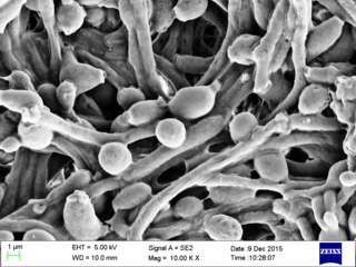
Candida albicans is an opportunistic pathogenic yeast that is a common member of the human gut flora. It can also survive outside the human body. It is detected in the gastrointestinal tract and mouth in 40–60% of healthy adults. It is usually a commensal organism, but it can become pathogenic in immunocompromised individuals under a variety of conditions. It is one of the few species of the genus Candida that causes the human infection candidiasis, which results from an overgrowth of the fungus. Candidiasis is, for example, often observed in HIV-infected patients. C. albicans is the most common fungal species isolated from biofilms either formed on (permanent) implanted medical devices or on human tissue. C. albicans, C. tropicalis, C. parapsilosis, and C. glabrata are together responsible for 50–90% of all cases of candidiasis in humans. A mortality rate of 40% has been reported for patients with systemic candidiasis due to C. albicans. By one estimate, invasive candidiasis contracted in a hospital causes 2,800 to 11,200 deaths yearly in the US. Nevertheless, these numbers may not truly reflect the true extent of damage this organism causes, given new studies indicating that C. albicans can cross the blood–brain barrier in mice.

<i>Cryptococcus neoformans</i> Species of yeast

Cryptococcus neoformans is an encapsulated yeast belonging to the class Tremellomycetes and an obligate aerobe that can live in both plants and animals. Its teleomorph is a filamentous fungus, formerly referred to Filobasidiella neoformans. In its yeast state, it is often found in bird excrement. Cryptococcus neoformans can cause disease in apparently immunocompetent, as well as immunocompromised, hosts.

Pathogenicity islands (PAIs), as termed in 1990, are a distinct class of genomic islands acquired by microorganisms through horizontal gene transfer. Pathogenicity islands are found in both animal and plant pathogens. Additionally, PAIs are found in both gram-positive and gram-negative bacteria. They are transferred through horizontal gene transfer events such as transfer by a plasmid, phage, or conjugative transposon. Therefore, PAIs contribute to microorganisms' ability to evolve.

<i>Aspergillus fumigatus</i> Species of fungus

Aspergillus fumigatus is a species of fungus in the genus Aspergillus, and is one of the most common Aspergillus species to cause disease in individuals with an immunodeficiency.

<i>Francisella tularensis</i> Species of bacterium

Francisella tularensis is a pathogenic species of Gram-negative coccobacillus, an aerobic bacterium. It is nonspore-forming, nonmotile, and the causative agent of tularemia, the pneumonic form of which is often lethal without treatment. It is a fastidious, facultative intracellular bacterium, which requires cysteine for growth. Due to its low infectious dose, ease of spread by aerosol, and high virulence, F. tularensis is classified as a Tier 1 Select Agent by the U.S. government, along with other potential agents of bioterrorism such as Yersinia pestis, Bacillus anthracis, and Ebola virus. When found in nature, Francisella tularensis can survive for several weeks at low temperatures in animal carcasses, soil, and water. In the laboratory, F. tularensis appears as small rods, and is grown best at 35–37 °C.

<i>Brucella suis</i> Bacterium that causes swine brucellosis

Brucella suis is a bacterium that causes swine brucellosis, a zoonosis that affects pigs. The disease typically causes chronic inflammatory lesions in the reproductive organs of susceptible animals or orchitis, and may even affect joints and other organs. The most common symptom is abortion in pregnant susceptible sows at any stage of gestation. Other manifestations are temporary or permanent sterility, lameness, posterior paralysis, spondylitis, and abscess formation. It is transmitted mainly by ingestion of infected tissues or fluids, semen during breeding, and suckling infected animals.

<i>Enterococcus faecalis</i> Species of bacterium

Enterococcus faecalis – formerly classified as part of the group D Streptococcus system – is a Gram-positive, commensal bacterium inhabiting the gastrointestinal tracts of humans. Like other species in the genus Enterococcus, E. faecalis is found in healthy humans and can be used as a probiotic. The probiotic strains such as Symbioflor1 and EF-2001 are characterized by the lack of specific genes related to drug resistance and pathogenesis. As an opportunistic pathogen, E. faecalis can cause life-threatening infections, especially in the nosocomial (hospital) environment, where the naturally high levels of antibiotic resistance found in E. faecalis contribute to its pathogenicity. E. faecalis has been frequently found in reinfected, root canal-treated teeth in prevalence values ranging from 30% to 90% of the cases. Re-infected root canal-treated teeth are about nine times more likely to harbor E. faecalis than cases of primary infections.

<i>Shigella flexneri</i> Species of bacterium

Shigella flexneri is a species of Gram-negative bacteria in the genus Shigella that can cause diarrhea in humans. Several different serogroups of Shigella are described; S. flexneri belongs to group B. S. flexneri infections can usually be treated with antibiotics, although some strains have become resistant. Less severe cases are not usually treated because they become more resistant in the future. Shigella are closely related to Escherichia coli, but can be differentiated from E.coli based on pathogenicity, physiology and serology.

<i>Yersinia pseudotuberculosis</i> Species of bacterium

Yersinia pseudotuberculosis is a Gram-negative bacterium that causes Far East scarlet-like fever in humans, who occasionally get infected zoonotically, most often through the food-borne route. Animals are also infected by Y. pseudotuberculosis. The bacterium is urease positive.

<i>Rhodococcus</i> Genus of bacteria

Rhodococcus is a genus of aerobic, nonsporulating, nonmotile Gram-positive bacteria closely related to Mycobacterium and Corynebacterium. While a few species are pathogenic, most are benign, and have been found to thrive in a broad range of environments, including soil, water, and eukaryotic cells. Some species have large genomes, including the 9.7 megabasepair genome of Rhodococcus sp. RHA1.

<i>Rhodococcus fascians</i> Species of bacterium

Rhodococcus fascians is a Gram positive bacterial phytopathogen that causes leafy gall disease. R. fascians is the only phytopathogenic member of the genus Rhodococcus; its host range includes both dicotyledonous and monocotyledonous hosts. Because it commonly afflicts tobacco (Nicotiana) plants, it is an agriculturally significant pathogen.

Listeriolysin O (LLO) is a hemolysin produced by the bacterium Listeria monocytogenes, the pathogen responsible for causing listeriosis. The toxin may be considered a virulence factor, since it is crucial for the virulence of L. monocytogenes.

<span class="mw-page-title-main">Virulence-related outer membrane protein family</span>

Virulence-related outer membrane proteins, or outer surface proteins (Osp) in some contexts, are expressed in the outer membrane of gram-negative bacteria and are essential to bacterial survival within macrophages and for eukaryotic cell invasion.

<i>Bacillus anthracis</i> Species of bacterium

Bacillus anthracis is a gram-positive and rod-shaped bacterium that causes anthrax, a deadly disease to livestock and, occasionally, to humans. It is the only permanent (obligate) pathogen within the genus Bacillus. Its infection is a type of zoonosis, as it is transmitted from animals to humans. It was discovered by a German physician Robert Koch in 1876, and became the first bacterium to be experimentally shown as a pathogen. The discovery was also the first scientific evidence for the germ theory of diseases.

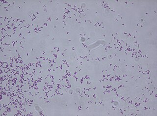
Streptococcus zooepidemicus is a Lancefield group C streptococcus that was first isolated in 1934 by P. R. Edwards, and named Animal pyogens A. It is a mucosal commensal and opportunistic pathogen that infects several animals and humans, but most commonly isolated from the uterus of mares. It is a subspecies of Streptococcus equi, a contagious upper respiratory tract infection of horses, and shares greater than 98% DNA homology, as well as many of the same virulence factors.

Enteroinvasive Escherichia coli (EIEC) is a type of pathogenic bacteria whose infection causes a syndrome that is identical to shigellosis, with profuse diarrhea and high fever. EIEC are highly invasive, and they use adhesin proteins to bind to and enter intestinal cells. They produce no toxins, but severely damage the intestinal wall through mechanical cell destruction.

Listeria ivanovii is a species of bacteria in the genus Listeria. The listeria are rod-shaped bacteria, do not produce spores, and become positively stained when subjected to Gram staining. Of the six bacteria species within the genus, L. ivanovii is one of the two pathogenic species. In 1955 Bulgaria, the first known isolation of this species was found from sheep. It behaves like L. monocytogenes, but is found almost exclusively in ruminants. The species is named in honor of Bulgarian microbiologist Ivan Ivanov. This species is facultatively anaerobic, which makes it possible for it to go through fermentation when there is oxygen depletion.

The alpha-D-phosphohexomutases are a large superfamily of enzymes, with members in all three domains of life. Enzymes from this superfamily are ubiquitous in organisms from E. Coli to humans, and catalyze a phosphoryl transfer reaction on a phosphosugar substrate. Four well studied subgroups in the superfamily are:

  1. Phosphoglucomutase (PGM)
  2. Phosphoglucomutase/Phosphomannomutase (PGM/PMM)
  3. Phosphoglucosamine mutase (PNGM)
  4. Phosphoaceytlglucosamine mutase (PAGM)
<i>Corynebacterium striatum</i> Species of bacterium

Corynebacterium striatum is a bacterium that is a member of the Corynebacterium genus. It is classified as non-diphtheritic. The bacterium is a gram-positive prokaryote that assumes a 'club-like' morphology, more formally known as a corynebacteria structure. It is non-lipophilic and undergoes aerobic respiration and is also a facultative anaerobe it is catalase negative and oxidase positive glucose and sucrose fermenter.

References

  1. Muscatello, G; Leadon, DP; Klayt, M; Ocampo-Sosa, A; Lewis, DA; Fogarty, U; Buckley, T; Gilkerson, JR; Meijer, WG; Vazquez-Boland, JA (September 2007). "Rhodococcus equi infection in foals: the science of 'rattles'". Equine Veterinary Journal. 39 (5): 470–8. doi:10.2746/042516407X209217. PMID   17910275.
  2. Makrai, L; Kobayashi, A; Matsuoka, M; Sasaki, Y; Kakuda, T; Dénes, B; Hajtós, I; Révész, I; Jánosi, K; Fodor, L; Varga, J; Takai, S (15 October 2008). "Isolation and characterisation of Rhodococcus equi from submaxillary lymph nodes of wild boars (Sus scrofa)". Veterinary Microbiology. 131 (3–4): 318–23. doi:10.1016/j.vetmic.2008.04.009. PMID   18499361.
  3. Kelly, B. G.; Wall, D. M.; Boland, C. A.; Meijer, W. G. (2002). "Isocitrate lyase of the facultative intracellular pathogen Rhodococcus equi". Microbiology. 148 (Pt 3): 793–798. doi: 10.1099/00221287-148-3-793 . PMID   11882714.
  4. von Bargen, K; Polidori, M; Becken, U; Huth, G; Prescott, JF; Haas, A (December 2009). "Rhodococcus equi virulence-associated protein A is required for diversion of phagosome biogenesis but not for cytotoxicity". Infection and Immunity. 77 (12): 5676–81. doi:10.1128/IAI.00856-09. PMC   2786453 . PMID   19797071.
  5. Fernandez-Mora, E; Polidori, M; Lührmann, A; Schaible, UE; Haas, A (August 2005). "Maturation of Rhodococcus equi-containing vacuoles is arrested after completion of the early endosome stage". Traffic. 6 (8): 635–53. doi: 10.1111/j.1600-0854.2005.00304.x . PMID   15998320. S2CID   30122137.
  6. Sydor, T; von Bargen, K; Hsu, FF; Huth, G; Holst, O; Wohlmann, J; Becken, U; Dykstra, T; Söhl, K; Lindner, B; Prescott, JF; Schaible, UE; Utermöhlen, O; Haas, A (March 2013). "Diversion of phagosome trafficking by pathogenic Rhodococcus equi depends on mycolic acid chain length". Cellular Microbiology. 15 (3): 458–73. doi:10.1111/cmi.12050. PMC   3864644 . PMID   23078612.
  7. Hondalus, MK; Mosser, DM (October 1994). "Survival and replication of Rhodococcus equi in macrophages". Infection and Immunity. 62 (10): 4167–75. doi:10.1128/IAI.62.10.4167-4175.1994. PMC   303092 . PMID   7927672.
  8. Lührmann, A; Mauder, N; Sydor, T; Fernandez-Mora, E; Schulze-Luehrmann, J; Takai, S; Haas, A (February 2004). "Necrotic death of Rhodococcus equi-infected macrophages is regulated by virulence-associated plasmids". Infection and Immunity. 72 (2): 853–62. doi:10.1128/iai.72.2.853-862.2004. PMC   321572 . PMID   14742529.
  9. Letek, M; Ocampo-Sosa, AA; Sanders, M; Fogarty, U; Buckley, T; Leadon, DP; González, P; Scortti, M; Meijer, WG; Parkhill, J; Bentley, S; Vázquez-Boland, JA (September 2008). "Evolution of the Rhodococcus equi vap pathogenicity island seen through comparison of host-associated vapA and vapB virulence plasmids". Journal of Bacteriology. 190 (17): 5797–805. doi:10.1128/JB.00468-08. PMC   2519538 . PMID   18606735.
  10. Takai, S; Hines, SA; Sekizaki, T; Nicholson, VM; Alperin, DA; Osaki, M; Takamatsu, D; Nakamura, M; Suzuki, K; Ogino, N; Kakuda, T; Dan, H; Prescott, JF (December 2000). "DNA sequence and comparison of virulence plasmids from Rhodococcus equi ATCC 33701 and 103". Infection and Immunity. 68 (12): 6840–7. doi:10.1128/iai.68.12.6840-6847.2000. PMC   97788 . PMID   11083803.
  11. Ren, J; Prescott, JF (1 July 2003). "Analysis of virulence plasmid gene expression of intra-macrophage and in vitro grown Rhodococcus equi ATCC 33701". Veterinary Microbiology. 94 (2): 167–82. doi:10.1016/S0378-1135(03)00099-3. PMID   12781484.
  12. Jain, S; Bloom, BR; Hondalus, MK (October 2003). "Deletion of vapA encoding Virulence Associated Protein A attenuates the intracellular actinomycete Rhodococcus equi". Molecular Microbiology. 50 (1): 115–28. doi: 10.1046/j.1365-2958.2003.03689.x . PMID   14507368. S2CID   42313934.
  13. Russell, DA; Byrne, GA; O'Connell, EP; Boland, CA; Meijer, WG (September 2004). "The LysR-type transcriptional regulator VirR is required for expression of the virulence gene vapA of Rhodococcus equi ATCC 33701". Journal of Bacteriology. 186 (17): 5576–84. doi:10.1128/JB.186.17.5576-5584.2004. PMC   516814 . PMID   15317761.
  14. 1 2 Garrity, GM (January 2014). "Conservation of Rhodococcus equi (Magnusson 1923) Goodfellow and Alderson 1977 and rejection of Corynebacterium hoagii (Morse 1912) Eberson 1918". International Journal of Systematic and Evolutionary Microbiology. 64 (Pt 1): 311–2. doi:10.1099/ijs.0.059741-0. PMID   24408953. Open Access logo PLoS transparent.svg
  15. Goodfellow, M; Sangal, V; Jones, AL; Sutcliffe, IC (September 2015). "Charting stormy waters: A commentary on the nomenclature of the equine pathogen variously named Prescottella equi, Rhodococcus equi and Rhodococcus hoagii". Equine Veterinary Journal. 47 (5): 508–509. doi: 10.1111/evj.12399 . PMID   25912143.
  16. 1 2 3 4 5 6 Berman, Jules J. (2012). Taxonomic guide to infectious diseases : understanding the biologic classes of pathogenic organisms. London: Elsevier/Academic Press. p. 266. ISBN   978-0-12-415895-5.

Further reading