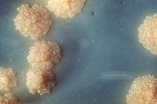
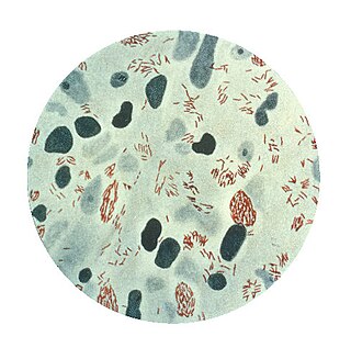
Mycobacterium tuberculosis, also known as Koch's bacillus, is a species of pathogenic bacteria in the family Mycobacteriaceae and the causative agent of tuberculosis. First discovered in 1882 by Robert Koch, M. tuberculosis has an unusual, waxy coating on its cell surface primarily due to the presence of mycolic acid. This coating makes the cells impervious to Gram staining, and as a result, M. tuberculosis can appear weakly Gram-positive. Acid-fast stains such as Ziehl–Neelsen, or fluorescent stains such as auramine are used instead to identify M. tuberculosis with a microscope. The physiology of M. tuberculosis is highly aerobic and requires high levels of oxygen. Primarily a pathogen of the mammalian respiratory system, it infects the lungs. The most frequently used diagnostic methods for tuberculosis are the tuberculin skin test, acid-fast stain, culture, and polymerase chain reaction.

Mycobacterium is a genus of over 190 species in the phylum Actinomycetota, assigned its own family, Mycobacteriaceae. This genus includes pathogens known to cause serious diseases in mammals, including tuberculosis and leprosy in humans. The Greek prefix myco- means 'fungus', alluding to this genus' mold-like colony surfaces. Since this genus has cell walls with Gram-positive and Gram-negative features, acid-fast staining is used to emphasize their resistance to acids, compared to other cell types.
Mycobacterium leprae, is one of the two species of bacteria that cause Hansen’s disease (leprosy), a chronic but curable infectious disease that damages the peripheral nerves and targets the skin, eyes, nose, and muscles.

Ethambutol is a medication primarily used to treat tuberculosis. It is usually given in combination with other tuberculosis medications, such as isoniazid, rifampicin and pyrazinamide. It may also be used to treat Mycobacterium avium complex, and Mycobacterium kansasii. It is taken by mouth.

Mycobacterium avium-intracellulare infection (MAI) is an atypical mycobacterial infection, i.e. one with nontuberculous mycobacteria or NTM, caused by Mycobacterium avium complex (MAC), which is made of two Mycobacterium species, M. avium and M. intracellulare. This infection causes respiratory illness in birds, pigs, and humans, especially in immunocompromised people. In the later stages of AIDS, it can be very severe. It usually first presents as a persistent cough. It is typically treated with a series of three antibiotics for a period of at least six months.
The Timpe and Runyon classification of nontuberculous mycobacteria based on the rate of growth, production of yellow pigment and whether this pigment was produced in the dark or only after exposure to light.
Mycobacterium celatum is a species of the phylum Actinomycetota, belonging to the genus Mycobacterium.
Mycobacterium goodii is an acid-fast bacterial species in the phylum Actinomycetota and the genus Mycobacterium.
Mycobacterium avium complex is a group of mycobacteria comprising Mycobacterium intracellulare and Mycobacterium avium that are commonly grouped because they infect humans together; this group, in turn, is part of the group of nontuberculous mycobacteria. These bacteria cause Mycobacterium avium-intracellulare infections or Mycobacterium avium complex infections in humans. These bacteria are common and are found in fresh and salt water, in household dust and in soil. MAC bacteria usually cause infection in those who are immunocompromised or those with severe lung disease.

Mycobacterium kansasii is a bacterium in the Mycobacterium genus. It is an environmental bacteria that causes opportunistic infections in humans, and is the one of the leading mycobacterial causes of human disease after tuberculosis and leprosy.
Mycobacterium wolinskyi is a rapidly growing mycobacterium most commonly seen in post-traumatic wound infections, especially those following open fractures and with associated osteomyelitis. Mycobacterium wolinskyi is clearly clinically significant, and occurs in the same settings as Mycobacterium smegmatis and members of the Mycobacterium fortuitum complex; they differ from members of the Mycobacterium fortuitum complex in the type of chronic lung disease they produce, with essentially all cases occurring in the setting of chronic lipoid pneumonia, either secondary to chronic oil ingestion or chronic aspiration. Etymology: Wolinsky, named after Emanuel Wolinsky in honour of, and in recognition for, significant contributions to the study of the non-tuberculous mycobacteria.
Mycobacterium tusciae is a slow-growing, scotochromogenic mycobacterium first isolated from a lymph node of an immunocompromised child and subsequently from tap water and from a respiratory specimen of a patient with chronic fibrosis. Etymology: tusciae referring to the Italian region of Tuscany, where the organisms were first isolated.
Mycobacteria that form colonies clearly visible to the naked eye in more than 7 days on subculture are termed slow growers.
Rapid growing mycobacterium consists of organism of the Mycobacterium fortuitum group and Mycobacterium chelonae/Mycobacterium abscessus group and these usually cause subcutaneous abscesses or cellulitis following trauma in immunocompetent patients.
The Xpert MTB/RIF is a cartridge-based nucleic acid amplification test (NAAT) for simultaneous rapid tuberculosis diagnosis and rapid antibiotic sensitivity test. It is an automated diagnostic test that can identify Mycobacterium tuberculosis (MTB) DNA and resistance to rifampicin (RIF). It was co-developed by the laboratory of Professor David Alland at the University of Medicine and Dentistry of New Jersey (UMDNJ), Cepheid Inc. and Foundation for Innovative New Diagnostics, with additional financial support from the US National Institutes of Health (NIH).
Mycobacterium parmense is a species of Mycobacterium.
Mycobacterium rhodesiae is a species of Mycobacterium.

Mycobacterium ulcerans is a species of bacteria found in various aquatic environments. The bacteria can infect humans and some other animals, causing persistent open wounds called Buruli ulcer. M. ulcerans is closely related to Mycobacterium marinum, from which it evolved around one million years ago, and more distantly to the mycobacteria which cause tuberculosis and leprosy.
Mycolicibacterium aichiense is a species of bacteria from the phylum Actinomycetota that was first isolated from soil and from human sputum. It produces pigments when grow in the dark and grows rapidly at 25–37 °C on Ogawa egg medium or Sauton agar medium.