
Leprosy, also known as Hansen's disease (HD), is a long-term infection by the bacteria Mycobacterium leprae or Mycobacterium lepromatosis. Infection can lead to damage of the nerves, respiratory tract, skin, and eyes. This nerve damage may result in a lack of ability to feel pain, which can lead to the loss of parts of a person's extremities from repeated injuries or infection through unnoticed wounds. An infected person may also experience muscle weakness and poor eyesight. Leprosy symptoms may begin within one year, but, for some people, symptoms may take 20 years or more to occur.

Mycobacterium is a genus of over 190 species in the phylum Actinomycetota, assigned its own family, Mycobacteriaceae. This genus includes pathogens known to cause serious diseases in mammals, including tuberculosis and leprosy in humans. The Greek prefix myco- means 'fungus', alluding to this genus' mold-like colony surfaces. Since this genus has cell walls with a waxy lipid-rich outer layer that contains high concentrations of mycolic acid, acid-fast staining is used to emphasize their resistance to acids, compared to other cell types.

A granuloma is an aggregation of macrophages that forms in response to chronic inflammation. This occurs when the immune system attempts to isolate foreign substances that it is otherwise unable to eliminate. Such substances include infectious organisms including bacteria and fungi, as well as other materials such as foreign objects, keratin, and suture fragments.
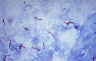
Mycobacterium leprae is one of the two species of bacteria that cause Hansen's disease (leprosy), a chronic but curable infectious disease that damages the peripheral nerves and targets the skin, eyes, nose, and muscles.

Erysipelothrix rhusiopathiae is a Gram-positive, catalase-negative, rod-shaped, non-spore-forming, nonacid-fast, nonmotile bacterium. Distributed worldwide, E. rhusiopathiae is primarily considered an animal pathogen, causing the disease known as erysipelas that may affect a wide range of animals. Pigs, turkeys and laying hens are most commonly affected, but cases have been reported in other mammals, birds, fish, and reptiles. In pigs, the disease is known as diamond skin disease. The bacterium can also cause zoonotic infections in humans, called erysipeloid. The human disease called erysipelas is not caused by E. rhusiopathiae, but by various members of the genus Streptococcus.

Buruli ulcer is an infectious disease characterized by the development of painless open wounds. The disease is limited to certain areas of the world, most cases occurring in Sub-Saharan Africa and Australia. The first sign of infection is a small painless nodule or area of swelling, typically on the arms or legs. The nodule grows larger over days to weeks, eventually forming an open ulcer. Deep ulcers can cause scarring of muscles and tendons, resulting in permanent disability.
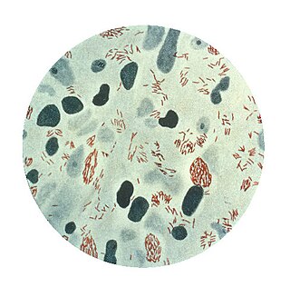
The Ziehl-Neelsen stain, also known as the acid-fast stain, is a bacteriological staining technique used in cytopathology and microbiology to identify acid-fast bacteria under microscopy, particularly members of the Mycobacterium genus. This staining method was initially introduced by Paul Ehrlich (1854–1915) and subsequently modified by the German bacteriologists Franz Ziehl (1859–1926) and Friedrich Neelsen (1854–1898) during the late 19th century.

Erythema nodosum (EN) is an inflammatory condition characterized by inflammation of the fat cells under the skin, resulting in tender red nodules or lumps that are usually seen on both shins. It can be caused by a variety of conditions, and typically resolves spontaneously within 30 days. It is common in young people aged 12–20 years.
Sporotrichosis, also known as rose handler's disease, is a fungal infection that may be localised to skin, lungs, bone and joint, or become systemic. It presents with firm painless nodules that later ulcerate. Following initial exposure to Sporothrix schenckii, the disease typically progresses over a period of a week to several months. Serious complications may develop in people who have a weakened immune system.

Mycobacteroides abscessus is a species of rapidly growing, multidrug-resistant, nontuberculous mycobacteria (NTM) that is a common soil and water contaminant. Although M. abscessus most commonly causes chronic lung infection and skin and soft tissue infection (SSTI), it can also cause infection in almost all human organs, mostly in patients with suppressed immune systems. Amongst NTM species responsible for disease, infection caused by M. abscessus complex are more difficult to treat due to antimicrobial drug resistance.
Mycobacterium celatum is a species of the phylum Actinomycetota, belonging to the genus Mycobacterium.
Mycobacterium duvalii is a species of the phylum Actinomycetota, belonging to the genus Mycobacterium.

Mycobacterium fortuitum is a nontuberculous species of the phylum Actinomycetota, belonging to the genus Mycobacterium.

Mycobacterium kansasii is a bacterium in the Mycobacterium genus. It is an environmental bacteria that causes opportunistic infections in humans, and is one of the leading mycobacterial causes of human disease after tuberculosis and leprosy.
Mycobacterium malmoense is a Gram-positive bacterium from the genus Mycobacterium.
Mycobacterium microti is a member of the Mycobacterium tuberculosis complex (MTBC) known as the 'Vole bacillus', first described as a pathogen of field voles in England.

Tuberculoid leprosy is a form of leprosy characterized by solitary skin lesions that are asymmetrically distributed with few lesions and well demarcated edges. There is also early and marked nerve damage. It tends to heal spontaneously. Tuberculoid leprosy is characterized by the formation of epithelioid cell granulomas consisting of a large number of epithelioid cells. In this form of leprosy, Mycobacterium leprae are either absent from the lesion or occur in very small numbers. This type of leprosy is the most benign and the least contagious.

Lepromatous leprosy is a form of leprosy characterized by pale macules in the skin.
Mycobacterium lepromatosis is an aerobic, acid-fast bacillus (AFB), and the second known causative agent of Hansen's disease (leprosy). It was discovered in 2008. Analysis of the 16S rRNA gene confirms that the species is distinct from Mycobacterium leprae.