Maltase-glucoamylase

Last updated
MGAM
PDB 2qly EBI.png
Available structures
PDB Human UniProt search: PDBe RCSB
Identifiers
Aliases MGAM , MG, MGA, Maltase-glucoamylase
External IDs OMIM: 154360 MGI: 1203495 HomoloGene: 130099 GeneCards: MGAM
Orthologs
SpeciesHumanMouse
Entrez
Ensembl
UniProt
RefSeq (mRNA)

NM_004668
NM_001365693

NM_001171003
NM_001368875

RefSeq (protein)

NP_004659
NP_001352622

n/a

Location (UCSC) Chr 7: 141.91 – 142.11 Mb Chr 6: 40.61 – 40.75 Mb
PubMed search [3] [4]
Wikidata
View/Edit Human View/Edit Mouse

Maltase-glucoamylase, intestinal is an enzyme that in humans is encoded by the MGAM gene. [5] [6]

Maltase-glucoamylase is an alpha-glucosidase digestive enzyme. It consists of two subunits with differing substrate specificity. Recombinant enzyme studies have shown that its N-terminal catalytic domain has highest activity against maltose, while the C-terminal domain has a broader substrate specificity and activity against glucose oligomers. [7] In the small intestine, this enzyme works in synergy with sucrase-isomaltase and alpha-amylase to digest the full range of dietary starches.

Gene

The MGAM gene –– which is located on chromosome 7q34 [8] –– codes for the protein Maltase-Glucoamylase. An alternative name for Maltase-Glucoamylase is glucan 1,4-alpha-glycosidase. [9]

Tissue distribution

Maltase-glucoamylase is a membrane-bound enzyme located in the intestinal walls. This lining of the intestine forms brush border in which food has to pass in order for the intestines to absorb the food. [10]

Enzymatic mechanism

This enzyme is a part of a family of enzymes called glycoside hydrolase family 31 (GH31). This is due to the digestive mechanism of the enzyme. GH31 enzymes undergo what is known as the Koshland double displacement mechanism [11] in which a glycosylation and deglycosylation step occurs, resulting in the retention of the overall configuration of the anomeric center. [12]

Structure

N-terminal maltase

The N-terminal maltase-glucoamylase enzymatic unit is in turn composed of 5 specific protein domains. The first of the 5 protein domains consist of a P-type trefoil domain [13] containing a cysteine rich domain. Second is an N-terminal beta-sandwich domain, identified via two antiparallel beta pleated sheets. The third and largest domain consists of a catalytic (beta/alpha) barrel type domain containing two inserted loops. The fourth and 5th domains are C-terminal domains, similar to the N-terminal beta-sandwich domain. The N-terminal Maltase-glucoamylase does not have the +2/+3 sugar binding active sites and so it cannot bind to larger substrates. The N-terminal domain shows its optimal enzymatic affinity for substrates maltose, maltotriose, maltotetrose, and maltopentose.

C-terminal glucase

The C-terminal glucase enzymatic unit contains extra binding sites, which allows for it to bind to larger substrates for catalytic digestion. [10] It was originally understood that maltase-glucoamylase's crystalline structure was inherently similar throughout the N and C-termini. Further studies have found that the C-terminus is composed of 21 more amino acid residues than the N-terminus, which account for its difference in function. Sucrase-Isomaltase –– located on chromosome 3q26–– has a similar crystalline structure to maltase-glucoamylase and work in tandem in the human small intestine. They have been derived from a common ancestor, as they both come from the same GH31 family. [8] As a result of having similar properties, both of these enzymes work together in the small intestine in order to convert consumed starch into glucose for metabolic energy. The difference between these two enzymes is that maltase-glucoamylase has a specific activity at the 1-4 linkage of sugar, where at SI has a specific activity at the 1-6 linkage. [10]

See also

Related Research Articles

<span class="mw-page-title-main">Lactase</span> Milk-sugar digesting enzyme

Lactase is an enzyme produced by many organisms and is essential to the complete digestion of whole milk. It breaks down the sugar lactose into its component parts, galactose and glucose. Lactase is found in the brush border of the small intestine of humans and other mammals. People deficient in lactase or lacking functional lactase may experience the symptoms of lactose intolerance after consuming milk products. Lactase can be purchased as a food supplement and is added to milk to produce "lactose-free" milk products.

<span class="mw-page-title-main">Small intestine</span> Organ in the gastrointestinal tract

The small intestine or small bowel is an organ in the gastrointestinal tract where most of the absorption of nutrients from food takes place. It lies between the stomach and large intestine, and receives bile and pancreatic juice through the pancreatic duct to aid in digestion. The small intestine is about 5.5 metres long and folds many times to fit in the abdomen. Although it is longer than the large intestine, it is called the small intestine because it is narrower in diameter.

<span class="mw-page-title-main">Maltase</span> Enzyme

Maltase is one type of alpha-glucosidase enzymes located in the brush border of the small intestine. This enzyme catalyzes the hydrolysis of disaccharide maltose into two simple sugars of glucose. Maltase is found in plants, bacteria, yeast, humans, and other vertebrates. It is thought to be synthesized by cells of the mucous membrane lining the intestinal wall.

<span class="mw-page-title-main">Acarbose</span> Chemical compound

Acarbose (INN) is an anti-diabetic drug used to treat diabetes mellitus type 2 and, in some countries, prediabetes. It is a generic sold in Europe and China as Glucobay, in North America as Precose, and in Canada as Prandase.

<span class="mw-page-title-main">Glycogen debranching enzyme</span> Mammalian protein found in Homo sapiens

The glycogen debranching enzyme, in humans, is the protein encoded by the gene AGL. This enzyme is essential for the breakdown of glycogen, which serves as a store of glucose in the body. It has separate glucosyltransferase and glucosidase activities.

Isomaltase is an enzyme that breaks the bonds linking saccharides, which cannot be broken by amylase or maltase. It digests polysaccharides at the alpha 1-6 linkages. Its substrate, alpha-limit dextrin, is a product of amylopectin digestion that retains its 1-6 linkage. The product of the enzymatic digestion of alpha-limit dextrin by isomaltase is maltose.

<span class="mw-page-title-main">Glycogen branching enzyme</span> Mammalian protein involved in glycogen production

1,4-alpha-glucan-branching enzyme, also known as brancher enzyme or glycogen-branching enzyme is an enzyme that in humans is encoded by the GBE1 gene.

Alpha-glucosidase inhibitors (AGIs) are oral anti-diabetic drugs used for diabetes mellitus type 2 that work by preventing the digestion of carbohydrates. Carbohydrates are normally converted into simple sugars (monosaccharides) by alpha-glucosidase enzymes present on cells lining the intestine, enabling monosaccharides to be absorbed through the intestine. Hence, alpha-glucosidase inhibitors reduce the impact of dietary carbohydrates on blood sugar.

α-Glucosidase Enzyme

α-Glucosidase (EC 3.2.1.20, is a glucosidase located in the brush border of the small intestine that acts upon α bonds:

<span class="mw-page-title-main">Trefoil knot fold</span>

The trefoil knot fold is a protein fold in which the protein backbone is twisted into a trefoil knot shape. "Shallow" knots in which the tail of the polypeptide chain only passes through a loop by a few residues are uncommon, but "deep" knots in which many residues are passed through the loop are extremely rare. Deep trefoil knots have been found in the SPOUT superfamily. including methyltransferase proteins involved in posttranscriptional RNA modification in all three domains of life, including bacterium Thermus thermophilus and proteins, in archaea and in eukaryota.

<span class="mw-page-title-main">Glycoside hydrolase</span> Class of enzymes which break glycosidic bonds via hydrolysis

In biochemistry, glycoside hydrolases are a class of enzymes which catalyze the hydrolysis of glycosidic bonds in complex sugars. They are extremely common enzymes, with roles in nature including degradation of biomass such as cellulose (cellulase), hemicellulose, and starch (amylase), in anti-bacterial defense strategies, in pathogenesis mechanisms and in normal cellular function. Together with glycosyltransferases, glycosidases form the major catalytic machinery for the synthesis and breakage of glycosidic bonds.

<span class="mw-page-title-main">Sucrose intolerance</span> Medical condition

Sucrose intolerance or genetic sucrase-isomaltase deficiency (GSID) is the condition in which sucrase-isomaltase, an enzyme needed for proper metabolism of sucrose (sugar) and starch, is not produced or the enzyme produced is either partially functional or non-functional in the small intestine. All GSID patients lack fully functional sucrase, while the isomaltase activity can vary from minimal functionality to almost normal activity. The presence of residual isomaltase activity may explain why some GSID patients are better able to tolerate starch in their diet than others with GSID.

<span class="mw-page-title-main">Sucrase-isomaltase</span>

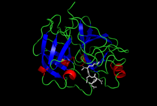
Sucrase-isomaltase is a bifunctional glucosidase located on the brush border of the small intestine, encoded by the human gene SI. It is a dual-function enzyme with two GH31 domains, one serving as the isomaltase, the other as a sucrose alpha-glucosidase. It has preferential expression in the apical membranes of enterocytes. The enzyme’s purpose is to digest dietary carbohydrates such as starch, sucrose and isomaltose. By further processing the broken-down products, energy in the form of ATP can be generated.

<span class="mw-page-title-main">GMP synthase</span>

Guanosine monophosphate synthetase, also known as GMPS is an enzyme that converts xanthosine monophosphate to guanosine monophosphate.

Glucan 1,4-α-glucosidase Enzyme that hydrolyses terminal α-1,4-D-glucose residues of polysaccharides

Glucan 1,4-α-glucosidase is an enzyme located on the brush border of the small intestine with systematic name 4-α-D-glucan glucohydrolase. It catalyses the following chemical reaction

<span class="mw-page-title-main">UDP-glucose 4-epimerase</span> Class of enzymes

The enzyme UDP-glucose 4-epimerase, also known as UDP-galactose 4-epimerase or GALE, is a homodimeric epimerase found in bacterial, fungal, plant, and mammalian cells. This enzyme performs the final step in the Leloir pathway of galactose metabolism, catalyzing the reversible conversion of UDP-galactose to UDP-glucose. GALE tightly binds nicotinamide adenine dinucleotide (NAD+), a co-factor required for catalytic activity.

<span class="mw-page-title-main">PRKCSH</span> Protein-coding gene in the species Homo sapiens

Glucosidase 2 subunit beta is an enzyme that in humans is encoded by the PRKCSH gene.

<span class="mw-page-title-main">Glycoside hydrolase family 31</span>

In molecular biology, glycoside hydrolase family 31 is a family of glycoside hydrolases.

Limit dextrinase is an enzyme with systematic name dextrin 6-alpha-glucanohydrolase. This enzyme catalyses the hydrolysis of (1->6)-alpha-D-glucosidic linkages in alpha- and beta-limits dextrins of amylopectin and glycogen, in amylopectin and pullulan.

Sucrose α-glucosidase is an enzyme with systematic name sucrose-α-D-glucohydrolase. It catalyses the hydrolysis of sucrose and maltose by an α-D-glucosidase-type action.

References

  1. 1 2 3 ENSG00000282607 GRCh38: Ensembl release 89: ENSG00000257335, ENSG00000282607 - Ensembl, May 2017
  2. 1 2 3 GRCm38: Ensembl release 89: ENSMUSG00000068587 - Ensembl, May 2017
  3. "Human PubMed Reference:". National Center for Biotechnology Information, U.S. National Library of Medicine.
  4. "Mouse PubMed Reference:". National Center for Biotechnology Information, U.S. National Library of Medicine.
  5. "Entrez Gene: maltase-glucoamylase (alpha-glucosidase)".
  6. Nichols BL, Eldering J, Avery S, Hahn D, Quaroni A, Sterchi E (January 1998). "Human small intestinal maltase-glucoamylase cDNA cloning. Homology to sucrase-isomaltase". The Journal of Biological Chemistry. 273 (5): 3076–81. doi: 10.1074/jbc.273.5.3076 . PMID   9446624.
  7. Quezada-Calvillo R, Sim L, Ao Z, Hamaker BR, Quaroni A, Brayer GD, et al. (April 2008). "Luminal starch substrate "brake" on maltase-glucoamylase activity is located within the glucoamylase subunit". The Journal of Nutrition. 138 (4): 685–92. doi: 10.1093/jn/138.4.685 . PMID   18356321.
  8. 1 2 Nichols BL, Avery S, Sen P, Swallow DM, Hahn D, Sterchi E (February 2003). "The maltase-glucoamylase gene: common ancestry to sucrase-isomaltase with complementary starch digestion activities". Proceedings of the National Academy of Sciences of the United States of America. 100 (3): 1432–7. Bibcode:2003PNAS..100.1432N. doi: 10.1073/pnas.0237170100 . PMC   298790 . PMID   12547908.
  9. Ao Z, Quezada-Calvillo R, Sim L, Nichols BL, Rose DR, Sterchi EE, Hamaker BR (May 2007). "Evidence of native starch degradation with human small intestinal maltase-glucoamylase (recombinant)". FEBS Letters. 581 (13): 2381–8. doi: 10.1016/j.febslet.2007.04.035 . PMID   17485087.
  10. 1 2 3 Sim L, Quezada-Calvillo R, Sterchi EE, Nichols BL, Rose DR (January 2008). "Human intestinal maltase-glucoamylase: crystal structure of the N-terminal catalytic subunit and basis of inhibition and substrate specificity". Journal of Molecular Biology. 375 (3): 782–92. doi:10.1016/j.jmb.2007.10.069. PMID   18036614.
  11. "Glycoside hydrolases". CAZypedia. Retrieved 2021-04-30.
  12. Frandsen TP, Svensson B (May 1998). "Plant alpha-glucosidases of the glycoside hydrolase family 31. Molecular properties, substrate specificity, reaction mechanism, and comparison with family members of different origin". Plant Molecular Biology. 37 (1): 1–13. doi:10.1023/A:1005925819741. PMID   9620260. S2CID   42054886.
  13. Otto B, Wright N (September 1994). "Trefoil peptides. Coming up clover". Current Biology. 4 (9): 835–8. doi:10.1016/S0960-9822(00)00186-X. PMID   7820556. S2CID   11245174.

Further reading