Related Research Articles

In all bilaterian animals, the mesoderm is one of the three primary germ layers in the very early embryo. The other two layers are the ectoderm and endoderm, with the mesoderm as the middle layer between them.

Embryology is the branch of biology that studies the prenatal development of gametes, fertilization, and development of embryos and fetuses. Additionally, embryology encompasses the study of congenital disorders that occur before birth, known as teratology.
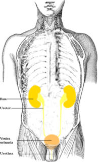
The ureters are tubes made of smooth muscle fibers that propel urine from the kidneys to the urinary bladder. In the human adult, the ureters are usually 25–30 cm (10–12 in) long and around 3–4 mm (0.12–0.16 in) in diameter. The ureter is lined by urothelial cells, a type of transitional epithelium, and has an additional smooth muscle layer in the more distal one-third to assist with peristalsis.

A morula is an early-stage embryo consisting of 16 cells in a solid ball contained within the zona pellucida.

Polyuria is excessive or an abnormally large production or passage of urine. Increased production and passage of urine may also be termed diuresis. Polyuria often appears in conjunction with polydipsia, though it is possible to have one without the other, and the latter may be a cause or an effect. Psychogenic polydipsia may lead to polyuria. Polyuria is usually viewed as a symptom or sign of another disorder, but it can be classed as a disorder, at least when its underlying causes are not clear.

The seminal vesicles, vesicular glands, or seminal glands, are a pair of simple tubular glands posteroinferior to the urinary bladder of some male mammals. Seminal vesicles are located within the pelvis. They secrete fluid that partly composes the semen.
In animal anatomy, a cloacakloh-AY-kə is the posterior orifice that serves as the only opening for the digestive, reproductive, and urinary tracts of many vertebrate animals, opening at the vent. All amphibians, reptiles, and a few mammals have this orifice, from which they excrete both urine and feces; this is in contrast to most placental mammals, which have two or three separate orifices for evacuation. Excretory openings with analogous purpose in some invertebrates are also sometimes referred to as cloacae. Mating by cloaca is known as cloacal copulation, mostly referred to as cloacal kiss.

The umbilical vein is a vein present during fetal development that carries oxygenated blood from the placenta into the growing fetus. The umbilical vein provides convenient access to the central circulation of a neonate for restoration of blood volume and for administration of glucose and drugs.

The paired submandibular glands are major salivary glands located beneath the floor of the mouth. They each weigh about 15 grams and contribute some 60–67% of unstimulated saliva secretion; on stimulation their contribution decreases in proportion as the parotid secretion rises to 50%.
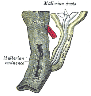
Paramesonephric ducts are paired ducts of the embryo that run down the lateral sides of the urogenital ridge and terminate at the sinus tubercle in the primitive urogenital sinus. In the female, they will develop to form the fallopian tubes, uterus, cervix, and the upper one-third of the vagina.
The male reproductive system consists of a number of sex organs that play a role in the process of human reproduction. These organs are located on the outside of the body and within the pelvis.

The anterior cerebral artery (ACA) is one of a pair of arteries on the brain that supplies oxygenated blood to most midline portions of the frontal lobes and superior medial parietal lobes. The two anterior cerebral arteries arise from the internal carotid artery and are part of the circle of Willis. The left and right anterior cerebral arteries are connected by the anterior communicating artery.
A myotome is the group of muscles that a single spinal nerve innervates. Similarly a dermatome is an area of skin that a single nerve innervates. In vertebrate embryonic development, a myotome is the part of a somite that develops into the muscles.

The tela choroidea is a region of meningeal pia mater and underlying ependyma that gives rise to the choroid plexus in each of the brain’s four ventricles. Tela is Latin for woven and is used to describe a web-like membrane or layer. The tela choroidea is a very thin part of the loose connective tissue of pia mater that overlies and closely adheres to the ependyma with no intervening tissue. It has a rich blood supply. The ependyma and vascular pia mater that make up the tela choroidea form regions of minute projections known as a choroid plexus that projects into each ventricle. The choroid plexus produces the cerebrospinal fluid of the ventricular system. The tela choroidea in the ventricles forms from different parts of the roof plate in the development of the embryo.
Neuroectoderm consists of cells derived from ectoderm. Formation of the neuroectoderm is first step in the development of the nervous system. The neuroectoderm receives bone morphogenetic protein-inhibiting signals from proteins such as noggin, which leads to the development of the nervous system from this tissue. Histologically, these cells are classified as pseudostratified columnar cells.
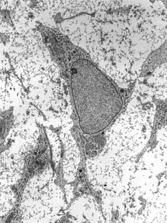
Mesenchyme is a type of connective tissue found mostly during embryonic development of bilateral animals (triploblasts). It is composed mainly of ground substance with few cells or fibers. It can also refer to a group of mucoproteins resembling mucus found, for example, in certain types of cysts. It is most easily found as a component of Wharton's jelly.

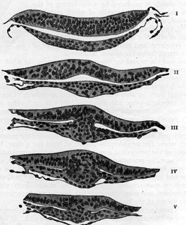
Human embryonic development, or human embryogenesis, refers to the development and formation of the human embryo. It is characterised by the process of cell division and cellular differentiation of the embryo that occurs during the early stages of development. In biological terms, the development of the human body entails growth from a one-celled zygote to an adult human being. Fertilisation occurs when the sperm cell successfully enters and fuses with an egg cell (ovum). The genetic material of the sperm and egg then combine to form a single cell called a zygote and the germinal stage of development commences. Embryonic development in the human, covers the first eight weeks of development; at the beginning of the ninth week the embryo is termed a fetus. Human embryology is the study of this development during the first eight weeks after fertilisation. The normal period of gestation (pregnancy) is ten months or 40 weeks.
The development of the reproductive system is a part of prenatal development, and concerns the sex organs. It is a part of the stages of sexual differentiation. Because its location, to a large extent, overlaps the urinary system, the development of them can also be described together as the development of the urinary and reproductive organs.
The development of the digestive system concerns the epithelium of the digestive system and the parenchyma of its derivatives, which originate from the endoderm. Connective tissue, muscular components, and peritoneal components originate in the mesoderm. Different regions of the gut tube such as the esophagus, stomach, duodenum, etc. are specified by a retinoic acid gradient that causes transcription factors unique to each region to be expressed. Differentiation of the gut and its derivatives depends upon reciprocal interactions between the gut endoderm and its surrounding mesoderm. Hox genes in the mesoderm are induced by a Hedgehog signaling pathway secreted by gut endoderm and regulate the craniocaudal organization of the gut and its derivatives. The gut system extends from the oropharyngeal membrane to the cloacal membrane and is divided into the foregut, midgut, and hindgut.
A cementicle is a small, spherical or ovoid calcified mass embedded within or attached to the cementum layer on the root surface of a tooth, or lying free within the periodontal ligament. They tend to occur in elderly individuals.
References
- ↑ Singh, Vishram Singh (2017). Textbook of Clinical Embryology (2 ed.). Elsevier Health Sciences. p. 511. ISBN 978-81-312-4882-9.
- ↑ Kelly, Christopher R.; Landman, Jaime, eds. (2012). The Netter Collection of Medical Illustrations: Urinary System. 5 (2 ed.). Elsevier Health Sciences. p. 30. ISBN 978-1-4377-2238-3.