Hydrocele

Last updated
Hydrocele
Other nameshydrocoele
Ultrasonography of hydrocele.jpg
Scrotal ultrasound of a 10 cm large hydrocele, with anechoic (dark) fluid surrounding the testicle
Specialty Urology

A hydrocele is an accumulation of serous fluid in a body cavity. A hydrocele testis, the most common form of hydrocele, is the accumulation of fluids around a testicle. It is often caused by fluid collecting within a layer wrapped around the testicle, called the tunica vaginalis, which is derived from peritoneum. Provided there is no hernia present, it goes away without treatment in the first year. Although hydroceles usually develop in males, rare instances have been described in females in the canal of Nuck. [1]

Contents

Primary hydroceles may develop in adulthood, particularly in the elderly and in hot countries, by slow accumulation of serous fluid. This is presumably caused by impaired reabsorption, which appears to be the explanation for most primary hydroceles, although the reason remains obscure.[ citation needed ] A hydrocele can also be the result of a plugged inguinal lymphatic system caused by repeated, chronic infection of Wuchereria bancrofti or Brugia malayi , two mosquito-borne parasites of Africa and Southeast Asia, respectively. As such, the condition would be a part of more diffuse sequelae commonly referred to as elephantiasis, which also affects the lymphatic system in other parts of the body.

Presentation

Complications

Among the complications of hydrocele are:[ citation needed ]

Complications are often diagnosed post-operatively, which can be differentiated through duplex ultrasound scanning and are bit observed until 24 to 48 hours for early complications such as drainage, infection, formation of haematocele, rupture, etc., but also for 1 to 6 weeks during follow-up on out-patient basis.[ citation needed ]

Cause

A hydrocele can be produced in four ways:

Primary hydroceles

The swelling is soft and non-tender, large in size on examination, and the testis cannot usually be felt. The presence of fluid is demonstrated by transillumination. These hydroceles can reach a huge size, containing large amount of fluid, as these are painless and are often ignored. They are otherwise asymptomatic, other than size and weight, causing inconvenience. However the long continued presence of large hydroceles causes atrophy of testis due to compression or by obstructing blood supply. In most cases, the hydrocele, when diagnosed early during complete physical examination, are small and the testis can easily be palpated within a lax hydrocele. However Ultrasound imaging is necessary to visualize the testis if the hydrocele sac is dense to reveal the primary abnormality. But these can become large in cases when left unattended. Hydroceles are usually painless, as are testicular tumors. A common method of diagnosing a hydrocele is by attempting to shine a strong light (transillumination) through the enlarged scrotum. A hydrocele will usually pass light, while a tumor will not (except in the case of a malignancy with reactive hydrocele).[ citation needed ]

Secondary hydroceles

Secondary hydroceles due to testicular diseases can be the result of cancer, trauma (such as a hernia), or orchitis (inflammation of testis), and can also occur in infants undergoing peritoneal dialysis. A hydrocele is not a cancer but it should be excluded clinically if a presence of a testicular tumor is suspected, however, there are no publications in the world literature that report a hydrocele in association with testicular cancer. Secondary hydrocele is most frequently associated with acute or chronic epididymo-orchitis. It is also seen with torsion of the testis and with some testicular tumors. A secondary hydrocele is usually lax and of moderate size: the underlying testis is palpable. A secondary hydrocele subsides when the primary lesion resolves.[ citation needed ]

Infantile hydroceles

It does not occur only in infants but it occurs in adults also, a hydrocele is usually an expression of a patent processus vaginalis (PPV). The tunica and the processus vaginalis are distended to the inguinal ring but there is no connection with the peritoneal cavity.[ citation needed ]

Congenital hydroceles

Communicating hydrocele, caused by the failure of the processus vaginalis closure. Ultrasound Scan ND 0124155309 1600360.png
Communicating hydrocele, caused by the failure of the processus vaginalis closure.

The processus vaginalis is patent and connects with the general peritoneal cavity. The communication is usually too small to allow herniation of intra-abdominal contents. Digital pressure on the hydrocele does not usually empty it, but the hydrocele fluid may drain into the peritoneal cavity when the child is lying down. Ascites or even ascitic tuberculous peritonitis should be considered if the swellings are bilateral.[ citation needed ]

Encysted hydrocele of the cord

There is a smooth oval swelling near the spermatic cord which is liable to be mistaken for an inguinal hernia. The swelling moves downwards and becomes less mobile if the testis is pulled gently downwards. Rarely, a hydrocele develops in a remnant of the processus vaginalis somewhere along the course of the spermatic cord. This hydrocele also transilluminates, and is known as an encysted hydrocele of the cord. In females, a related region in females, a multicystic hydrocele of the canal of Nuck [3] sometimes presents as a swelling in the groin. It probably results from cystic degeneration of the round ligament. Unlike a hydrocele of the cord, a hydrocele of the canal of Nuck is always at least partially within the inguinal canal.[ citation needed ]

The accuracy of the diagnosis must be ascertained. Great care must be taken to differentiate a hydrocele from a scrotal hernia or tumor of the testicle. Ultrasound imaging can be very useful in these cases. A hernia usually can be reduced, transmits a cough impulse, and is not translucent. A hydrocele cannot be reduced into the inguinal canal and gives no impulse on coughing unless a hernia is also present. In young children a hydrocele is often associated with a complete congenital type of hernial sac.[ citation needed ]

Diagnosis

A primary hydrocele is described as having the following characteristics:[ citation needed ]

Treatment

Most hydroceles appearing in the first year of life resolve without treatment. [4] Hydroceles that persist after the first year or occur later in life require treatment only in selected cases, such as patients who are symptomatic with pain or a pressure sensation, or when the scrotal skin integrity is compromised from chronic irritation; the treatment of choice is surgery and the operation is conducted via an open access technique aiming to excise the hydrocele sac. [5] [6] Anesthesia is required for the operation; general anesthesia is of choice in children, while spinal anesthesia is usually sufficient in adults. Local infiltration anesthesia is not satisfactory because it cannot abolish abdominal pain due to traction on the spermatic cord. [7] In longstanding cases, hydrocele fluid may be opalescent with cholesterol and may contain crystals of tyrosine. [8]

After aspiration of a primary hydrocele, fluid reaccumulates over the following months and periodic aspiration or operation is needed. For younger patients, operation is usually preferred.[ citation needed ] Sclerotherapy is an alternative; after aspiration, 6% aqueous phenol (10–20 ml) together with 1% lidocaine for analgesia can be injected, and this often inhibits reaccumulation. These alternative treatments are generally regarded as unsatisfactory because of the high incidence of recurrences and the frequent necessity for repetition of the procedure.[ citation needed ]

Related Research Articles

<span class="mw-page-title-main">Testicle</span> Internal organ in the male reproductive system

A testicle or testis is the male reproductive gland or gonad in all bilaterians, including humans. It is homologous to the female ovary. The functions of the testes are to produce both sperm and androgens, primarily testosterone. Testosterone release is controlled by the anterior pituitary luteinizing hormone, whereas sperm production is controlled both by the anterior pituitary follicle-stimulating hormone and gonadal testosterone.

<span class="mw-page-title-main">Testicular torsion</span> Medical condition

Testicular torsion occurs when the spermatic cord twists, cutting off the blood supply to the testicle. The most common symptom in children is sudden, severe testicular pain. The testicle may be higher than usual in the scrotum and vomiting may occur. In newborns, pain is often absent and instead the scrotum may become discolored or the testicle may disappear from its usual place.

<span class="mw-page-title-main">Testicular cancer</span> Medical condition

Testicular cancer is cancer that develops in the testicles, a part of the male reproductive system. Symptoms may include a lump in the testicle or swelling or pain in the scrotum. Treatment may result in infertility.

<span class="mw-page-title-main">Spermatic cord</span> Structure in the human male reproductive system

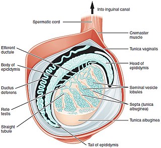
The spermatic cord is the cord-like structure in males formed by the vas deferens and surrounding tissue that runs from the deep inguinal ring down to each testicle. Its serosal covering, the tunica vaginalis, is an extension of the peritoneum that passes through the transversalis fascia. Each testicle develops in the lower thoracic and upper lumbar region and migrates into the scrotum. During its descent it carries along with it the vas deferens, its vessels, nerves etc. There is one on each side.

<span class="mw-page-title-main">Inguinal canal</span> Human abdominal anatomy

The inguinal canal is a passage in the anterior abdominal wall on each side of the body which in males convey the spermatic cords and in females the round ligament of the uterus. The inguinal canals are larger and more prominent in males.

<span class="mw-page-title-main">Hydrocele testis</span> Medical condition

A hydrocele testis is an accumulation of clear fluid within the cavum vaginale, the potential space between the layers of the tunica vaginalis of the testicle. It is the most common form of hydrocele and is often referred to simply as a "hydrocele". A primary hydrocele testis causes a painless enlargement in the scrotum on the affected side and is thought to be due to the defective absorption of fluid secreted between the two layers of the tunica vaginalis. A secondary hydrocele is secondary to either inflammation or a neoplasm in the testis.

Orchitis is inflammation of the testes. It can also involve swelling, pains and frequent infection, particularly of the epididymis, as in epididymitis. The term is from the Ancient Greek ὄρχις meaning "testicle"; same root as orchid.

<span class="mw-page-title-main">Orchiopexy</span> Surgery to fix a testicle into the scrotum

Orchiopexy is a surgery to move and/or permanently fix a testicle into the scrotum. While orchiopexy typically describes the operation to surgically correct an undescended testicle, it is also used to resolve testicular torsion.

<span class="mw-page-title-main">Inguinal hernia</span> Medical condition in which contents of the abdominal cavity protrude through the inguinal canal

An inguinal hernia or groin hernia, is a hernia (protrusion) of abdominal cavity contents through the inguinal canal. Symptoms, which may include pain or discomfort especially with or following coughing, exercise, or bowel movements, are absent in about a third of patients. Symptoms often get worse throughout the day and improve when lying down. A bulging area may occur that becomes larger when bearing down. Inguinal hernias occur more often on the right than left side. The main concern is strangulation, where the blood supply to part of the intestine is blocked. This usually produces severe pain and tenderness of the area.

<span class="mw-page-title-main">Spermatocele</span> Medical condition

Spermatocele is a fluid-filled cyst that develops in the epididymis. The fluid is usually a clear or milky white color and may contain sperm. Spermatoceles are typically filled with spermatozoa and they can vary in size from several millimeters to many centimeters. Small spermatoceles are relatively common, occurring in an estimated 30 percent of males. They are generally not painful. However, some people may experience discomfort such as a dull pain in the scrotum from larger spermatoceles. They are not cancerous, nor do they cause an increased risk of testicular cancer. Additionally, unlike varicoceles, they do not reduce fertility.

<span class="mw-page-title-main">Hematocele</span> Medical condition

A hematocele is a collections of blood in a body cavity or potential space. The term most commonly refers to the collection of blood in the tunica vaginalis around the testes, known as a scrotal hematocele. Hematoceles can also occur in the abdominal cavity and other body cavities. Hematoceles are rare, making them harder to diagnose and treat. They are very common especially as slowly growing masses in the scrotum usually in men older than 50 years.

Transillumination is the technique of sample illumination by transmission of light through the sample. Transillumination is used in a variety of methods of imaging.

Inguinal orchiectomy is a specific method of orchiectomy whereby one or both testicles and the full spermatic cord are surgically removed through an incision in the lower lateral abdomen. The procedure is generally performed by a urologist, typically if testicular cancer is suspected. Often it is performed as same-day surgery, with the patient returning home within hours of the procedure. Some patients elect to have a prosthetic testicle inserted into their scrotum. Depending on whether or not a prosthetic testicle is put in place of the original one, operating times run on average from three to six hours.

The canal of Nuck, first described by Anton Nuck in 1691, is an abnormal patent (open) pouch of peritoneum extending into the labia majora of women. It is analogous to a patent processus vaginalis in males. In rare cases, it may give rise to a cyst or a hydrocele in women and has potential to develop into an indirect inguinal hernia. The pouch accompanies the gubernaculum during development of the urinary and reproductive organs, more specifically during the descent of the ovaries, and normally obliterates.

<span class="mw-page-title-main">Tunica vaginalis</span> Pouch of serous membrane that covers the testes

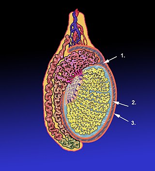
The tunica vaginalis is a pouch of serous membrane within the scrotum that lines the testis and epididymis, and the inner surface of the scrotum. It is the outermost of the three layers that constitute the capsule of the testis, with the tunica albuginea of penis situated beneath it.

<span class="mw-page-title-main">Vaginal process</span> An embryonic developmental outpouching of the parietal peritoneum

The vaginal process is an embryonic developmental outpouching of the parietal peritoneum. It is present from around the 12th week of gestation, and commences as a peritoneal outpouching.

The development of the gonads is part of the prenatal development of the reproductive system and ultimately forms the testes in males and the ovaries in females. The gonads initially develop from the mesothelial layer of the peritoneum.

<span class="mw-page-title-main">Scrotum</span> Sac of skin that protects the testicles

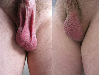
The scrotum or scrotal sac is an anatomical male reproductive structure located at the base of the penis that consists of a suspended dual-chambered sac of skin and smooth muscle. It is present in most terrestrial male mammals. The scrotum contains the external spermatic fascia, testicles, epididymis, and ductus deferens. It is a distention of the perineum and carries some abdominal tissues into its cavity including the testicular artery, testicular vein, and pampiniform plexus. The perineal raphe is a small, vertical, slightly raised ridge of scrotal skin under which is found the scrotal septum. It appears as a thin longitudinal line that runs front to back over the entire scrotum. In humans and some other mammals, the scrotum becomes covered with pubic hair at puberty. The scrotum will usually tighten during penile erection and when exposed to cold temperatures. One testis is typically lower than the other to avoid compression in the event of an impact.

<span class="mw-page-title-main">Scrotal ultrasound</span> Medical ultrasound examination of the scrotum.

Scrotalultrasound is a medical ultrasound examination of the scrotum. It is used in the evaluation of testicular pain, and can help identify solid masses.

Male genital examination is a physical examination of the genital in males to detect ailments and to assess sexual development and is normally included in an annual physical examination. The examination includes checking the penis, scrotum, and urethral meatus. A comprehensive assessment of the male genitals assesses the pubic hair based on Sexual Maturity Rating and the size of the testicles and penis. The exam is conducted to detect various ailments, measure sexual development, or verify a person's age and biological sex. The genitourinary system can also be assessed as part of the male genital examination. During a genital examination, the doctor can detect any of the following: structural abnormalities, urethral opening abnormalities, problems related to uncircumcision, lumps, tumors, redness, excoriation, edema, lesions, swelling, cancer, hair-related issues and many others. In some instances where a physical examination of the male genitals is not sufficient to diagnose an individual than an internal genital examination using imaging or ultrasounds will be needed for further evaluation.

References

  1. Sarkar, Santanu; Panja, Soumyajyoti; Kumar, Sandeep (February 2016). "Hydrocele of the Canal of Nuck (Female Hydrocele): A Rare Differential for Inguino-Labial Swelling". Journal of Clinical and Diagnostic Research. 10 (2): PD21–PD22. doi:10.7860/JCDR/2016/16710.7284. ISSN   2249-782X. PMC   4800595 . PMID   27042529.
  2. Bailey & Love's/24th/1407-1409
  3. "hydrocele of the canal of Nuck" . GPnotebook.
  4. "Hydrocele: Symptoms, causes, and treatment". www.medicalnewstoday.com. 2020-03-24. Retrieved 2020-12-17.
  5. Zollinger's Atlas of Surgeries
  6. "UpToDate". www.uptodate.com. Retrieved 2020-01-06.
  7. Manual of Anesthesia for Out Patient Surgical Procedures
  8. Bailey and Love-Short Practice of Surgery