Related Research Articles

A skeleton is the structural frame that supports the body of most animals. There are several types of skeletons, including the exoskeleton, which is a rigid outer shell that holds up an organism's shape; the endoskeleton, a rigid internal frame to which the organs and soft tissues attach; and the hydroskeleton, a flexible internal structure supported by the hydrostatic pressure of body fluids.

In vertebrate anatomy, ribs are the long curved bones which form the rib cage, part of the axial skeleton. In most tetrapods, ribs surround the chest, enabling the lungs to expand and thus facilitate breathing by expanding the chest cavity. They serve to protect the lungs, heart, and other internal organs of the thorax. In some animals, especially snakes, ribs may provide support and protection for the entire body.

The maxilla in vertebrates is the upper fixed bone of the jaw formed from the fusion of two maxillary bones. In humans, the upper jaw includes the hard palate in the front of the mouth. The two maxillary bones are fused at the intermaxillary suture, forming the anterior nasal spine. This is similar to the mandible, which is also a fusion of two mandibular bones at the mandibular symphysis. The mandible is the movable part of the jaw.

The vomer is one of the unpaired facial bones of the skull. It is located in the midsagittal line, and articulates with the sphenoid, the ethmoid, the left and right palatine bones, and the left and right maxillary bones. The vomer forms the inferior part of the nasal septum in humans, with the superior part formed by the perpendicular plate of the ethmoid bone. The name is derived from the Latin word for a ploughshare and the shape of the bone.

Aquatic respiration is the process whereby an aquatic organism exchanges respiratory gases with water, obtaining oxygen from oxygen dissolved in water and excreting carbon dioxide and some other metabolic waste products into the water.

The quadrate bone is a skull bone in most tetrapods, including amphibians, sauropsids, and early synapsids.
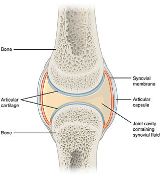
A synovial joint, also known as diarthrosis, joins bones or cartilage with a fibrous joint capsule that is continuous with the periosteum of the joined bones, constitutes the outer boundary of a synovial cavity, and surrounds the bones' articulating surfaces. This joint unites long bones and permits free bone movement and greater mobility. The synovial cavity/joint is filled with synovial fluid. The joint capsule is made up of an outer layer of fibrous membrane, which keeps the bones together structurally, and an inner layer, the synovial membrane, which seals in the synovial fluid.
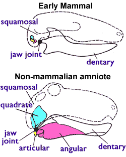
The articular bone is part of the lower jaw of most vertebrates, including most jawed fish, amphibians, birds and various kinds of reptiles, as well as ancestral mammals.
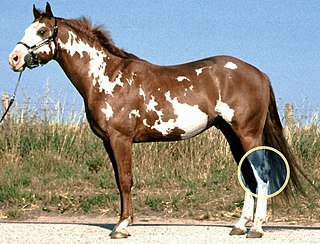
The hock, or gambrel, is the joint between the tarsal bones and tibia of a digitigrade or unguligrade quadrupedal mammal, such as a horse, cat, or dog. This joint may include articulations between tarsal bones and the fibula in some species, while in others the fibula has been greatly reduced and is only found as a vestigial remnant fused to the distal portion of the tibia. It is the anatomical homologue of the ankle of the human foot. While homologous joints occur in other tetrapods, the term is generally restricted to mammals, particularly long-legged domesticated species.

Endochondral ossification is one of the two essential processes during fetal development of the mammalian skeletal system by which bone tissue is produced. Unlike intramembranous ossification, the other process by which bone tissue is produced, cartilage is present during endochondral ossification. Endochondral ossification is also an essential process during the rudimentary formation of long bones, the growth of the length of long bones, and the natural healing of bone fractures.

The pharyngeal arches, also known as visceral arches, are structures seen in the embryonic development of vertebrates that are recognisable precursors for many structures. In fish, the arches are known as the branchial arches, or gill arches.

In humans, the cartilaginous bar of the mandibular arch is formed by what are known as Meckel's cartilages also known as Meckelian cartilages; above this the incus and malleus are developed. Meckel's cartilage arises from the first pharyngeal arch.

The evolution of mammalian auditory ossicles was an evolutionary process that resulted in the formation of the bones of the mammalian middle ear. These bones, or ossicles, are a defining characteristic of all mammals. The event is well-documented and important as a demonstration of transitional forms and exaptation, the re-purposing of existing structures during evolution.

In human anatomy, the neurocranium, also known as the braincase, brainpan, or brain-pan is the upper and back part of the skull, which forms a protective case around the brain. In the human skull, the neurocranium includes the calvaria or skullcap. The remainder of the skull is the facial skeleton.

The endocranium in comparative anatomy is a part of the skull base in vertebrates and it represents the basal, inner part of the cranium. The term is also applied to the outer layer of the dura mater in human anatomy.
The parasphenoid is a bone which can be found in the cranium of many vertebrates. It is an unpaired dermal bone which lies at the midline of the roof of the mouth. In many reptiles, it fuses to the endochondral (cartilage-derived) basisphenoid bone of the lower braincase, forming a bone known as the parabasisphenoid. Early mammals have a small parasphenoid, but for the most part its function has been replaced by the vomer bone. The parasphenoid has been lost in placental mammals and caecilian amphibians.

The sternum or breastbone is a long flat bone located in the central part of the chest. It connects to the ribs via cartilage and forms the front of the rib cage, thus helping to protect the heart, lungs, and major blood vessels from injury. Shaped roughly like a necktie, it is one of the largest and longest flat bones of the body. Its three regions are the manubrium, the body, and the xiphoid process. The word sternum originates from Ancient Greek στέρνον (stérnon) 'chest'.
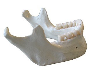
In anatomy, the mandible, lower jaw or jawbone is the bottom skeleton that makes up the lower half of the mouth in jawed vertebrates. In arthropods, the largest pair of appendages of their mouthparts are also named mandibles.

The vertebral column, also known as the backbone or spine, is the core part of the axial skeleton in vertebrate animals. The vertebral column is the defining characteristic of vertebrate endoskeleton in which the notochord found in all chordates has been replaced by a segmented series of mineralized irregular bones called vertebrae, separated by fibrocartilaginous intervertebral discs. The dorsal portion of the vertebral column houses the spinal canal, a cavity formed by alignment of the neural arches that encloses and protects the spinal cord.
References
- ↑ Wilkins, Adam S. (2017). "How the Face Develops". Making Faces. Harvard University Press. p. 56. ISBN 9780674725522.
- 1 2 Kent, George C.; Carr, Robert K. (2001). Comparative Anatomy of the Vertebrates (9th ed.). New York, NY: McGraw-Hill. ISBN 0-07-303869-5.
| Authority control databases: National |
|---|
| | This anatomy article is a stub. You can help Wikipedia by expanding it. |