Mesencephalic locomotor region

Last updated
Mesencephalic locomotor region
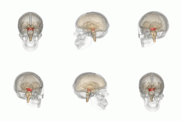
Midbrain small.gif
Details
Part of Brainstem
Identifiers
Acronym(s)MLR
Anatomical terminology

The mesencephalic locomotor region (MLR) is a functionally defined area of the midbrain that is associated with the initiation and control of locomotor movements in vertebrate species. [1] [2]

Contents

Neuroanatomical organization

The MLR was first described by Shik and colleagues in 1966 when they observed that electrical stimulation of a region of the midbrain in decerebrate cats produced walking and running behavior on a treadmill. [3] Twenty-eight years later, Masdeu and colleagues described the presence of a MLR in humans. [4] It is now widely acknowledged that, along with other motor control centers of the brain, the MLR plays an active role in initiating and modulating the spinal neural circuitry to control posture and gait. [5] Anatomically, as the name suggests, the MLR is located in the mesencephalon (or midbrain), ventral to the inferior colliculus and near the cuneiform nucleus. [6] Although identifying the exact anatomical substrates of the MLR has been subject to considerable debate, the pedunculopontine nucleus (PPN), cuneiform nucleus, and midbrain extrapyramidal area are thought to form the neuroanatomical basis of the MLR. [7] [8] [9] Nuclei within the MLR receive inputs from the substantia nigra of the basal ganglia and neural centers within the limbic system. [10] Projections from the MLR descend via the medullary and pontine reticulospinal tracts to act on spinal motor neurons supplying the trunk and proximal limb flexors and extensors. [2] [5] [11]

The PPN within the MLR is composed of a diverse population of neurons containing the neurotransmitters gamma-amino-butyric acid (GABA), glutamate, and acetylcholine (ACh). [12] Results from animal and clinical studies suggest that cholinergic neurons in the PPN play a crucial role in modulating both the rhythm of locomotion and postural muscle tone. [13] [14] Glutamatergic and cholinergic inputs from the MLR may be responsible for regulating the excitability of reticulospinal neurons that in turn project to spinal central pattern generators to initiate stepping. [1] [15]

Clinical significance

The integration of motor and sensory information during walking involves communication between cortical, subcortical, and spinal circuits. Step-like motor patterns of the lower extremities can be induced through activation of the spinal circuitry alone; [16] however, supraspinal input is necessary for functional bipedal walking in humans. [17] [18] Pathologies of the nuclei within the MLR have been associated with a combination of clinical features that are unique to midbrain dysfunction and can be differentiated from other subcortical neurological conditions such as those associated with Parkinsonism and cerebellar lesions. [19]

In a clinical case series, three adult males with isolated lesions of the MLR presented with gait hesitation and gait ataxia characterized by stepping that lacked uniform direction, amplitude, and rhythmicity. [20] Although gait hesitation and ataxia are also clinical features of Parkinson's disease and lesions of the cerebellum, respectively, the authors noted that the patients did not display any other common signs or symptoms associated with these neurological conditions, suggesting that pathologies of the midbrain can produce gait disturbances even when cerebellar and basal ganglia function are intact. In a study investigating high-level gait and balance disorders in elderly adults who had no evidence of rheumatologic, orthopedic, or neurologic disease, brain imaging data revealed an association between reduced gray matter density of the PPN and cuneiform nucleus and impaired gait initiation, step execution, and postural control. [21] Additionally, among eighteen individuals with Parkinson's disease who either did or did not experience Freezing of Gait, functional magnetic resonance imaging revealed reduced activity in the MLR and supplementary motor area among those individuals who experienced episodic gait hesitation. [22] Freezing of Gait has also been associated with functional reorganization of supraspinal locomotor networks whereby altered connectivity and communication between the supplementary motor area and MLR were observed. [23] These findings suggest that the MLR does in fact play a unique role in human locomotion, especially with respect to step initiation and motor planning.

Deep brain stimulation

Given the role of the MLR in gait initiation and postural control, researchers and clinicians have investigated the effects of targeted deep brain stimulation (DBS) on gait disturbances in clinical populations. [24] [25] Plaha and Gill reported significant improvements in gait dysfunction and postural instability in two patients with advanced Parkinson's disease who were treated using DBS electrodes implanted in the region of the PPN. [26] Likewise, in a more recent study, six patients with Parkinson's disease demonstrated improvements in posture, gait, and postural stability following 6 months of DBS to the PPN and subthalamic nucleus. [27] Bachmann and colleagues applied DBS to the MLR in rats with chronic, incomplete spinal cord injury and reported improved hindlimb function and near normal restoration of locomotor function following treatment. [28]

See also

Related Research Articles

<span class="mw-page-title-main">Central nervous system</span> Brain and spinal cord

The central nervous system (CNS) is the part of the nervous system consisting of the brain and spinal cord, the retina and optic nerve, and the olfactory nerve and epithelia. The CNS is so named because the brain integrates the received information and coordinates and influences the activity of all parts of the bodies of bilaterally symmetric and triploblastic animals—that is, all multicellular animals except sponges and diploblasts. It is a structure composed of nervous tissue positioned along the rostral to caudal axis of the body and may have an enlarged section at the rostral end which is a brain. Only arthropods, cephalopods and vertebrates have a true brain, though precursor structures exist in onychophorans, gastropods and lancelets.

<span class="mw-page-title-main">Deep brain stimulation</span> Neurosurgical treatment involving implantation of a brain pacemaker

Deep brain stimulation (DBS) is a surgical procedure that implants a neurostimulator and electrodes which sends electrical impulses to specified targets in the brain responsible for movement control. The treatment is designed for a range of movement disorders such as Parkinson's disease, essential tremor, and dystonia, as well as for certain neuropsychiatric conditions like obsessive-compulsive disorder (OCD) and epilepsy. The exact mechanisms of DBS are complex and not entirely clear, but it is known to modify brain activity in a structured way.

<span class="mw-page-title-main">Extrapyramidal system</span> Connection between brain and spinal cord

In anatomy, the extrapyramidal system is a part of the motor system network causing involuntary actions. The system is called extrapyramidal to distinguish it from the tracts of the motor cortex that reach their targets by traveling through the pyramids of the medulla. The pyramidal tracts may directly innervate motor neurons of the spinal cord or brainstem, whereas the extrapyramidal system centers on the modulation and regulation of anterior (ventral) horn cells.

<span class="mw-page-title-main">Gait (human)</span> A pattern of limb movements made during locomotion

A gait is a manner of limb movements made during locomotion. Human gaits are the various ways in which humans can move, either naturally or as a result of specialized training. Human gait is defined as bipedal forward propulsion of the center of gravity of the human body, in which there are sinuous movements of different segments of the body with little energy spent. Varied gaits are characterized by differences such as limb movement patterns, overall velocity, forces, kinetic and potential energy cycles, and changes in contact with the ground.

<span class="mw-page-title-main">Subthalamic nucleus</span> Small lens-shaped nucleus in the brain

The subthalamic nucleus (STN) is a small lens-shaped nucleus in the brain where it is, from a functional point of view, part of the basal ganglia system. In terms of anatomy, it is the major part of the subthalamus. As suggested by its name, the subthalamic nucleus is located ventral to the thalamus. It is also dorsal to the substantia nigra and medial to the internal capsule. It was first described by Jules Bernard Luys in 1865, and the term corpus Luysi or Luys' body is still sometimes used.

<span class="mw-page-title-main">Reticular formation</span> Spinal trigeminal nucleus

The reticular formation is a set of interconnected nuclei that are located in the brainstem, hypothalamus, and other regions. It is not anatomically well defined, because it includes neurons located in different parts of the brain. The neurons of the reticular formation make up a complex set of networks in the core of the brainstem that extend from the upper part of the midbrain to the lower part of the medulla oblongata. The reticular formation includes ascending pathways to the cortex in the ascending reticular activating system (ARAS) and descending pathways to the spinal cord via the reticulospinal tracts.

<span class="mw-page-title-main">Pontine tegmentum</span>

The pontine tegmentum, or dorsal pons, is located within the brainstem, and is one of two parts of the pons, the other being the ventral pons or basilar part of the pons. The pontine tegmentum can be defined in contrast to the basilar pons: basilar pons contains the corticospinal tract running craniocaudally and can be considered the rostral extension of the ventral medulla oblongata; however, basilar pons is distinguished from ventral medulla oblongata in that it contains additional transverse pontine fibres that continue laterally to become the middle cerebellar peduncle. The pontine tegmentum is all the material dorsal from the basilar pons to the fourth ventricle. Along with the dorsal surface of the medulla, it forms part of the rhomboid fossa – the floor of the fourth ventricle.

<span class="mw-page-title-main">Hypokinesia</span> Decreased movement due to basal ganglia dysfunction

Hypokinesia is one of the classifications of movement disorders, and refers to decreased bodily movement. Hypokinesia is characterized by a partial or complete loss of muscle movement due to a disruption in the basal ganglia. Hypokinesia is a symptom of Parkinson's disease shown as muscle rigidity and an inability to produce movement. It is also associated with mental health disorders and prolonged inactivity due to illness, amongst other diseases.

The pedunculopontine nucleus (PPN) or pedunculopontine tegmental nucleus is a collection of neurons located in the upper pons in the brainstem. It lies caudal to the substantia nigra and adjacent to the superior cerebellar peduncle. It has two divisions of subnuclei; the pars compacta containing mainly cholinergic neurons, and the pars dissipata containing mainly glutamatergic neurons and some non-cholinergic neurons. The pedunculopontine nucleus is one of the main components of the reticular activating system. It was first described in 1909 by Louis Jacobsohn-Lask, a German neuroanatomist.

Central pattern generators (CPGs) are self-organizing biological neural circuits that produce rhythmic outputs in the absence of rhythmic input. They are the source of the tightly-coupled patterns of neural activity that drive rhythmic and stereotyped motor behaviors like walking, swimming, breathing, or chewing. The ability to function without input from higher brain areas still requires modulatory inputs, and their outputs are not fixed. Flexibility in response to sensory input is a fundamental quality of CPG-driven behavior. To be classified as a rhythmic generator, a CPG requires:

  1. "two or more processes that interact such that each process sequentially increases and decreases, and
  2. that, as a result of this interaction, the system repeatedly returns to its starting condition."

The zona incerta (ZI) is a horizontally elongated region of gray matter in the subthalamus below the thalamus. Its connections project extensively over the brain from the cerebral cortex down into the spinal cord.

In the management of Parkinson's disease, due to the chronic nature of Parkinson's disease (PD), a broad-based program is needed that includes patient and family education, support-group services, general wellness maintenance, exercise, and nutrition. At present, no cure for the disease is known, but medications or surgery can provide relief from the symptoms.

<span class="mw-page-title-main">Mesencephalic nucleus of trigeminal nerve</span>

The mesencephalic nucleus of trigeminal nerve is one of the sensory nuclei of the trigeminal nerve. It is located in the brainstem. It receives proprioceptive sensory information from the muscles of mastication and other muscles of the head and neck. It is involved in processing information about the position of the jaw/teeth. It is functionally responsible for preventing excessive biting that may damage the dentition, regulating tooth pain perception, and mediating the jaw jerk reflex.

<span class="mw-page-title-main">Internal globus pallidus</span>

The internal globus pallidus and the external globus pallidus (GPe) make up the globus pallidus. The GPi is one of the output nuclei of the basal ganglia. The GABAergic neurons of the GPi send their axons to the ventral anterior nucleus (VA) and the ventral lateral nucleus (VL) in the dorsal thalamus, to the centromedian complex, and to the pedunculopontine complex.

<span class="mw-page-title-main">Blocq's disease</span> Loss of memory of specialized movements causing the inability to maintain an upright posture

Blocq's disease was first considered by Paul Blocq (1860–1896), who described this phenomenon as the loss of memory of specialized movements causing the inability to maintain an upright posture, despite normal function of the legs in the bed. The patient is able to stand up, but as soon as the feet are on the ground, the patient cannot hold himself upright nor walk; however when lying down, the subject conserved the integrity of muscular force and the precision of movements of the lower limbs. The motivation of this study came when a fellow student Georges Marinesco (1864) and Paul published a case of parkinsonian tremor (1893) due to a tumor located in the substantia nigra.

<span class="mw-page-title-main">Parkinsonian gait</span> Type of gait due to Parkinsons disease

Parkinsonian gait is the type of gait exhibited by patients with Parkinson's disease (PD). It is often described by people with Parkinson's as feeling like being stuck in place, when initiating a step or turning, and can increase the risk of falling. This disorder is caused by a deficiency of dopamine in the basal ganglia circuit leading to motor deficits. Gait is one of the most affected motor characteristics of this disorder although symptoms of Parkinson's disease are varied.

<span class="mw-page-title-main">Neural substrate of locomotor central pattern generators in mammals</span>

Central pattern generators are biological neural networks organized to produce any rhythmic output without requiring a rhythmic input. In mammals, locomotor CPGs are organized in the lumbar and cervical segments of the spinal cord, and are used to control rhythmic muscle output in the arms and legs. Certain areas of the brain initiate the descending neural pathways that ultimately control and modulate the CPG signals. In addition to this direct control, there exist different feedback loops that coordinate the limbs for efficient locomotion and allow for the switching of gaits under appropriate circumstances.

Gait variability seen in Parkinson's Disorders arise due to cortical changes induced by pathophysiology of the disease process. Gait rehabilitation is focused to harness the adapted connections involved actively to control these variations during the disease progression. Gait variabilities seen are attributed to the defective inputs from the Basal Ganglia. However, there is altered activation of other cortical areas that support the deficient control to bring about a movement and maintain some functional mobility.

A descending neuron is a neuron that conveys signals from the brain to neural circuits in the spinal cord (vertebrates) or ventral nerve cord (invertebrates). As the sole conduits of information between the brain and the body, descending neurons play a key role in behavior. Their activity can initiate, maintain, modulate, and terminate behaviors such as locomotion. Because the number of descending neurons is several orders of magnitude smaller than the number of neurons in either the brain or spinal cord/ventral nerve cord, this class of cells represents a critical bottleneck in the flow of information from sensory systems to motor circuits.

<span class="mw-page-title-main">Interlimb coordination</span> Coordination of the left and right limbs

Interlimb coordination is the coordination of the left and right limbs. It could be classified into two types of action: bimanual coordination and hands or feet coordination. Such coordination involves various parts of the nervous system and requires a sensory feedback mechanism for the neural control of the limbs. A model can be used to visualize the basic features, the control centre of locomotor movements, and the neural control of interlimb coordination. This coordination mechanism can be altered and adapted for better performance during locomotion in adults and for the development of motor skills in infants. The adaptive feature of interlimb coordination can also be applied to the treatment for CNS damage from stroke and the Parkinson's disease in the future.

References

  1. 1 2 Le Ray, D; Juvin, L; Ryczko, D; Dubuc, R (2011). "Chapter 4 - Supraspinal control of locomotion: the mesencephalic locomotor region" (PDF). Progress in Brain Research. 188: 51–70. doi:10.1016/B978-0-444-53825-3.00009-7. PMID   21333802.
  2. 1 2 Pahapill, P; Lozano, A (2000). "The pedunculopontine nucleus and Parkinson's disease". Brain. 123 (9): 1767–1783. doi: 10.1093/brain/123.9.1767 . PMID   10960043.
  3. Shik, ML; Severin, FV; Orlofsky, GN (1966). "Control of walking and running by means of electrical stimulation of the midbrain". Biophysics (Oxf). 11: 756–765.
  4. Masdeu, JC; Alampur, U; Cavaliere, R; Tavoulareas, G (1994). "Astasia and gait failure with damage of the pontomesencephalic locomotor region". Annals of Neurology. 35 (5): 619–621. doi:10.1002/ana.410350517. PMID   8179307. S2CID   2193366.
  5. 1 2 Takakusaki, K (2017). "Functional neuroanatomy for posture and gait control". Journal of Movement Disorders. 10 (1): 1–17. doi:10.14802/jmd.16062. PMC   5288669 . PMID   28122432.
  6. Pearson, KG; Gordon, JE (2013). Principles of Neural Science: Locomotion (5th ed.). New York: The McGraw-Hill Companies, Inc.
  7. Skinner, RD; Garcia-Rill, E (1984). "The mesencephalic locomotor region (MLR) in the rat". Brain Research. 323 (2): 385–389. doi:10.1016/0006-8993(84)90319-6. PMID   6525525. S2CID   46258649.
  8. Chang, Stephano J.; Cajigas, Iahn; Opris, Ioan; Guest, James D.; Noga, Brian R. (2020-08-21). "Dissecting Brainstem Locomotor Circuits: Converging Evidence for Cuneiform Nucleus Stimulation". Frontiers in Systems Neuroscience. 14: 64. doi: 10.3389/fnsys.2020.00064 . ISSN   1662-5137. PMC   7473103 . PMID   32973468.
  9. Sherman, D; Fuller, PM; Marcus, J; Yu, J; Zhang, P; Chamberlin, NL; Saper, CB; Lu, J (2015). "Anatomical location of the mesencephalic locomotor region and its possible role in locomotion, posture, cataplexy, and Parkinsonism". Frontiers in Neurology. 6 (140): 140. doi: 10.3389/fneur.2015.00140 . PMC   4478394 . PMID   26157418.
  10. Sherman, D; Fuller, PM; Marcus, J; Yu, J; Zhang, P; Chamberlin, N; Saper, C; Lu, J (2015). "Anatomical location of the mesencephalic locomotor region and its possible role in locomotion, posture, cataplexy, and Parkinsonism". Frontiers in Neurology. 6 (140): 140. doi: 10.3389/fneur.2015.00140 . PMC   4478394 . PMID   26157418.
  11. Takakusaki, K; Tomita, N; Yano, M (2008). "Substrates for normal gait and pathophysiology of gait disturbances with respect to the basal ganglia dysfunction". Journal of Neurology. 255 (Suppl 4): 19–29. doi:10.1007/s00415-008-4004-7. PMID   18821082. S2CID   22009992.
  12. Takakusaki, K; Chiba, R; Tsukasa, N; Okumura, T (2016). "Brainstem control of locomotion and muscle tone with special reference to the role of the mesopontine tegmentum and medullary reticulospinal systems". Journal of Neural Transmission. 123 (7): 695–729. doi:10.1007/s00702-015-1475-4. PMC   4919383 . PMID   26497023.
  13. Bohnen, NI; Albin, RL (2011). "The cholinergic system and Parkinson disease". Behavioural Brain Research. 221 (2): 564–573. doi:10.1016/j.bbr.2009.12.048. PMC   2888997 . PMID   20060022.
  14. Takakusaki, K; Obara, K; Nozu, T; Okumura, T (2011). "Modulatory effects of the GABAergic basal ganglia neurons on the PPN and the muscle tone inhibitory system in cats". Archives Italiennes de Biologie. 149 (4): 385–405. doi:10.4449/aib.v149i4.1383. PMID   22205597.
  15. Skinner, RD; Kinjo, N; Henderson, V; Garcia-Rill, E (1990). "Locomotor projections from the pedunculopontine nucleus to the spinal cord". Neurological Reports. 1 (3): 183–186. doi:10.1097/00001756-199011000-00001. PMID   2129877.
  16. Whelan, PJ (2003). "Developmental aspects of spinal locomotor function: insights from using the in vitro mouse spinal cord preparation". Journal of Physiology. 553 (Pt 3): 695–706. doi:10.1113/jphysiol.2003.046219. PMC   2343637 . PMID   14528025.
  17. Nielsen, Jens Bo (2003). "How we walk: central control of muscle activity during human walking". Neuroscientist. 9 (3): 195–204. doi:10.1177/1073858403009003012. PMID   15065815. S2CID   34448912.
  18. Capaday, Charles (2002). "The special nature of human walking and its neural control". Trends in Neurosciences. 25 (7): 370–376. doi:10.1016/s0166-2236(02)02173-2. PMID   12079766. S2CID   19997715.
  19. Ruchalski, K; Hathout, GM (2012). "A medley of midbrain maladies: a brief review of midbrain anatomy and syndromology for radiologists". Radiology Research and Practice. 2012: 258524. doi: 10.1155/2012/258524 . PMC   3366251 . PMID   22693668.
  20. Hathout, GM; Bhidayasiri, R (2005). "Midbrain ataxia: an introduction to the mesencephalic locomotor region and the pedunculopontine nucleus". American Journal of Roentgenology. 184 (3): 953–956. doi:10.2214/ajr.184.3.01840953. PMID   15728623.
  21. Demain, A; Westby, M; Fernandez-Vidal, S; Karachi, C; Bonneville, F; Do, MC; Delmaire, C; Dormont, D; Bardinet, E; Agid, Y; Chastan, N; Welter, ML (2014). "High-level gait and balance disorders in the elderly: a midbrain disease?". Journal of Neurology. 261 (1): 196–206. doi:10.1007/s00415-013-7174-x. PMC   3895186 . PMID   24202784.
  22. Peterson, DS; Pickett, KA; Duncan, R; Perlmutter, J; Earhart, GM (2014). "Gait-related brain activity in people with Parkinson disease with freezing of gait". PLOS ONE. 9 (3): e90634. Bibcode:2014PLoSO...990634P. doi: 10.1371/journal.pone.0090634 . PMC   3940915 . PMID   24595265.
  23. Fling, BW; Cohen, RG; Mancini, M; Carpenter, SD; Fair, DA; Nutt, JG; Horak, FB (2014). "Functional reorganization of the locomotor network in Parkinson patients with freezing gait". PLOS ONE. 9 (6): e100291. Bibcode:2014PLoSO...9j0291F. doi: 10.1371/journal.pone.0100291 . PMC   4061081 . PMID   24937008.
  24. Hamani, C; Scellig, S; Laxton, A; Lozano, AM (2007). "The pedunculopontine nucleus and movement disorders: anatomy and the role for deep brain stimulation". Parkinsonism and Related Disorders. 13: S276-80. doi:10.1016/s1353-8020(08)70016-6. PMID   18267250.
  25. Richardson, M (2014). "Deep brain stimulation for locomotor recovery following spinal cord injury". Neurosurgery. 74 (2): N18-9. doi: 10.1227/01.neu.0000442979.07078.ac . PMID   24435148.
  26. Plaha, P; Gill, S (2005). "Bilateral deep brain stimulation of the pedunculopontine nucleus for Parkinson's disease". NeuroReport. 16 (17): 1883–1887. doi:10.1097/01.wnr.0000187637.20771.a0. PMID   16272872. S2CID   20912030.
  27. Stefani, A; Lozano, AM; Peppe, A; Stanzione, P; Galati, S; Tropepi, D (2007). "Bilateral deep brain stimulation of the pedunculopontine and subthalamic nuclei in severe Parkinson's disease" (PDF). Brain. 130 (6): 1596–1607. doi: 10.1093/brain/awl346 . PMID   17251240.
  28. Bachmann, LC; Matis, A; Lindau, NT; Felder, P; Gullo, M; Schwab, ME (2013). "Deep brain stimulation of the midbrain locomotor region improves paretic hindlimb function after spinal cord injury in rats". Science Translational Medicine. 5 (208): 208. doi:10.1126/scitranslmed.3005972. PMID   24154600. S2CID   39733797.