Internal globus pallidus

Last updated
Internal globus pallidus
Anatomie-Basalganglien-A.jpg
Internal globus pallidus (GPi) is seen in the 2nd image from the left
Details
Part of Globus pallidus
Identifiers
Latin globus pallidus internus,
globus pallidus medialis
Acronym(s)GPi
NeuroNames 233
NeuroLex ID birnlex_1555
TA98 A14.1.09.511
TA2 5572
FMA 61840
Anatomical terms of neuroanatomy

The internal globus pallidus (GPi or medial globus pallidus; in rodents its homologue is known as the entopeduncular nucleus) and the external globus pallidus (GPe) make up the globus pallidus. The GPi is one of the output nuclei of the basal ganglia (the other being the substantia nigra pars reticulata). The GABAergic neurons of the GPi send their axons to the ventral anterior nucleus (VA) and the ventral lateral nucleus (VL) in the dorsal thalamus, to the centromedian complex, and to the pedunculopontine complex. [1] [2]

Contents

The efferent bundle is constituted first of the ansa and lenticular fasciculus, then crosses the internal capsule within and in parallel to the Edinger's comb system then arrives at the laterosuperior corner of the subthalamic nucleus and constitutes the field H2 of Forel, then H, and suddenly changes its direction to form field H1 that goes to the inferior part of the thalamus. The distribution of axonal islands is widespread in the lateral region of the thalamus. The innervation of the central region is done by collaterals. [3]

The internal globus pallidus contains GABAergic neurons, which allow for its inhibitory function. As the GPi, along with the substantia nigra pars reticulata, forms the output of the basal ganglia, these neurons extend to the thalamus, the centromedian complex and the pedunculopontine complex. [4]

Function

Direct and indirect striatopallidal pathways: Glutamatergic pathways are red, dopaminergic are magenta and GABAergic pathways are blue. STN: Subthalamic Nucleus SNr: Substantia Nigra pars reticulata SNc: Substantia Nigra pars compacta GPe: External globus pallidus Basal-ganglia-classic.png
Direct and indirect striatopallidal pathways: Glutamatergic pathways are red, dopaminergic are magenta and GABAergic pathways are blue. STN: Subthalamic Nucleus SNr: Substantia Nigra pars reticulata SNc: Substantia Nigra pars compacta GPe: External globus pallidus

The GPi acts to tonically inhibit the ventral lateral nucleus and ventral anterior nucleus of the thalamus. As these two nuclei are needed for movement planning, this inhibition restricts movement initiation and prevents unwanted movements.

Direct pathway

The GPi receives inhibitory GABAergic signals from the striatum by way of striatopallidal fibres, when a movement requirement is signaled from the cerebral cortex. As the GPi is one of the direct output centers of the basal ganglia, this causes disinhibition of the thalamus, increasing overall ease of initiating and maintaining movement. As this pathway only contains one synapse (from the striatum to the internal globus pallidus), it is known as the direct pathway. [5]

The direct pathway is modulated by stimulation of the GPi by the external globus pallidus and subthalamic nucleus, via the indirect pathway. [6]

Clinical significance

Dysfunction of the internal globus pallidus has been correlated to Parkinson's disease, [7] Tourette syndrome, [8] and tardive dyskinesia. [9]

The internal globus pallidus is the target of deep brain stimulation (DBS) for these diseases. Deep brain stimulation sends regulated electrical pulses to the target. In patients with tardive dyskinesia treated with DBS, most people reported more than a 50% improvement in symptoms. [9] Tourette syndrome patients have also benefited from this treatment, showing over 50% improvement in tic severity (compulsive disabling motor tics are symptoms of Tourette patients). [8] The GPi is also considered a "highly effective target for neuromodulation" when using deep brain stimulation on Parkinson's disease patients. [7] There is seen to be only some involvement in Huntington's disease [10] with mostly the external globus pallidus being affected. [11]

Related Research Articles

<span class="mw-page-title-main">Putamen</span> Round structure at the base of the forebrain

The putamen is a round structure located at the base of the forebrain (telencephalon). The putamen and caudate nucleus together form the dorsal striatum. It is also one of the structures that compose the basal nuclei. Through various pathways, the putamen is connected to the substantia nigra, the globus pallidus, the claustrum, and the thalamus, in addition to many regions of the cerebral cortex. A primary function of the putamen is to regulate movements at various stages and influence various types of learning. It employs GABA, acetylcholine, and enkephalin to perform its functions. The putamen also plays a role in degenerative neurological disorders, such as Parkinson's disease.

<span class="mw-page-title-main">Striatum</span> Nucleus in the basal ganglia of the brain

The striatum or corpus striatum is a nucleus in the subcortical basal ganglia of the forebrain. The striatum is a critical component of the motor and reward systems; receives glutamatergic and dopaminergic inputs from different sources; and serves as the primary input to the rest of the basal ganglia.

<span class="mw-page-title-main">Substantia nigra</span> Structure in the basal ganglia of the brain

The substantia nigra (SN) is a basal ganglia structure located in the midbrain that plays an important role in reward and movement. Substantia nigra is Latin for "black substance", reflecting the fact that parts of the substantia nigra appear darker than neighboring areas due to high levels of neuromelanin in dopaminergic neurons. Parkinson's disease is characterized by the loss of dopaminergic neurons in the substantia nigra pars compacta.

<span class="mw-page-title-main">Basal ganglia</span> Group of subcortical nuclei involved in the motor and reward systems

The basal ganglia (BG) or basal nuclei are a group of subcortical nuclei found in the brains of vertebrates. In humans and other primates, differences exist, primarily in the division of the globus pallidus into external and internal regions, and in the division of the striatum. Positioned at the base of the forebrain and the top of the midbrain, they have strong connections with the cerebral cortex, thalamus, brainstem and other brain areas. The basal ganglia are associated with a variety of functions, including regulating voluntary motor movements, procedural learning, habit formation, conditional learning, eye movements, cognition, and emotion.

<span class="mw-page-title-main">Deep brain stimulation</span> Neurosurgical treatment involving implantation of a brain pacemaker

Deep brain stimulation (DBS) is a surgical procedure that implants a neurostimulator and electrodes which sends electrical impulses to specified targets in the brain responsible for movement control. The treatment is designed for a range of movement disorders such as Parkinson's disease, essential tremor, and dystonia, as well as for certain neuropsychiatric conditions like obsessive-compulsive disorder (OCD) and epilepsy. The exact mechanisms of DBS are complex and not entirely clear, but it is known to modify brain activity in a structured way.

<span class="mw-page-title-main">Globus pallidus</span> Structure of the basal ganglia of the brain

The globus pallidus (GP), also known as paleostriatum or dorsal pallidum, is a subcortical structure of the brain. It consists of two adjacent segments, one external, known in rodents simply as the globus pallidus, and one internal, known in rodents as the entopeduncular nucleus. It is part of the telencephalon, but retains close functional ties with the subthalamus in the diencephalon – both of which are part of the extrapyramidal motor system. The globus pallidus is a major component of the basal ganglia, with principal inputs from the striatum, and principal direct outputs to the thalamus and the substantia nigra. The latter is made up of similar neuronal elements, has similar afferents from the striatum, similar projections to the thalamus, and has a similar synaptology. Neither receives direct cortical afferents, and both receive substantial additional inputs from the intralaminar thalamus.

<span class="mw-page-title-main">Dopaminergic pathways</span> Projection neurons in the brain that synthesize and release dopamine

Dopaminergic pathways in the human brain are involved in both physiological and behavioral processes including movement, cognition, executive functions, reward, motivation, and neuroendocrine control. Each pathway is a set of projection neurons, consisting of individual dopaminergic neurons.

<span class="mw-page-title-main">Nigrostriatal pathway</span> Bilateral pathway in the brain

The nigrostriatal pathway is a bilateral dopaminergic pathway in the brain that connects the substantia nigra pars compacta (SNc) in the midbrain with the dorsal striatum in the forebrain. It is one of the four major dopamine pathways in the brain, and is critical in the production of movement as part of a system called the basal ganglia motor loop. Dopaminergic neurons of this pathway release dopamine from axon terminals that synapse onto GABAergic medium spiny neurons (MSNs), also known as spiny projection neurons (SPNs), located in the striatum.

<span class="mw-page-title-main">Indirect pathway</span> Neuronal circuit that suppresses unwanted movements

The indirect pathway, sometimes known as the indirect pathway of movement, is a neuronal circuit through the basal ganglia and several associated nuclei within the central nervous system (CNS) which helps to prevent unwanted muscle contractions from competing with voluntary movements. It operates in conjunction with the direct pathway.

<span class="mw-page-title-main">Subthalamic nucleus</span> Small lens-shaped nucleus in the brain

The subthalamic nucleus (STN) is a small lens-shaped nucleus in the brain where it is, from a functional point of view, part of the basal ganglia system. In terms of anatomy, it is the major part of the subthalamus. As suggested by its name, the subthalamic nucleus is located ventral to the thalamus. It is also dorsal to the substantia nigra and medial to the internal capsule. It was first described by Jules Bernard Luys in 1865, and the term corpus Luysi or Luys' body is still sometimes used.

<span class="mw-page-title-main">Hyperkinesia</span> Excessive movements due to basal ganglia dysfunction

Hyperkinesia refers to an increase in muscular activity that can result in excessive abnormal movements, excessive normal movements, or a combination of both. Hyperkinesia is a state of excessive restlessness which is featured in a large variety of disorders that affect the ability to control motor movement, such as Huntington's disease. It is the opposite of hypokinesia, which refers to decreased bodily movement, as commonly manifested in Parkinson's disease.

<span class="mw-page-title-main">Hypokinesia</span> Decreased movement due to basal ganglia dysfunction

Hypokinesia is one of the classifications of movement disorders, and refers to decreased bodily movement. Hypokinesia is characterized by a partial or complete loss of muscle movement due to a disruption in the basal ganglia. Hypokinesia is a symptom of Parkinson's disease shown as muscle rigidity and an inability to produce movement. It is also associated with mental health disorders and prolonged inactivity due to illness, amongst other diseases.

The zona incerta (ZI) is a horizontally elongated region of gray matter in the subthalamus below the thalamus. Its connections project extensively over the brain from the cerebral cortex down into the spinal cord.

<span class="mw-page-title-main">Primate basal ganglia</span>

The basal ganglia form a major brain system in all species of vertebrates, but in primates there are special features that justify a separate consideration. As in other vertebrates, the primate basal ganglia can be divided into striatal, pallidal, nigral, and subthalamic components. In primates, however, there are two pallidal subdivisions called the external globus pallidus (GPe) and internal globus pallidus (GPi). Also in primates, the dorsal striatum is divided by a large tract called the internal capsule into two masses named the caudate nucleus and the putamen—in most other species no such division exists, and only the striatum as a whole is recognized. Beyond this, there is a complex circuitry of connections between the striatum and cortex that is specific to primates. This complexity reflects the difference in functioning of different cortical areas in the primate brain.

<span class="mw-page-title-main">Medium spiny neuron</span> Type of GABAergic neuron in the striatum

Medium spiny neurons (MSNs), also known as spiny projection neurons (SPNs), are a special type of GABAergic inhibitory cell representing 95% of neurons within the human striatum, a basal ganglia structure. Medium spiny neurons have two primary phenotypes : D1-type MSNs of the direct pathway and D2-type MSNs of the indirect pathway. Most striatal MSNs contain only D1-type or D2-type dopamine receptors, but a subpopulation of MSNs exhibit both phenotypes.

<span class="mw-page-title-main">External globus pallidus</span> Part of the globus pallidus

The external globus pallidus combines with the internal globus pallidus (GPi) to form the globus pallidus, an anatomical subset of the basal ganglia. Globus pallidus means "pale globe" in Latin, indicating its appearance. The external globus pallidus is the segment of the globus pallidus that is relatively further (lateral) from the midline of the brain.

<span class="mw-page-title-main">Basal ganglia disease</span> Group of physical problems resulting from basal ganglia dysfunction

Basal ganglia disease is a group of physical problems that occur when the group of nuclei in the brain known as the basal ganglia fail to properly suppress unwanted movements or to properly prime upper motor neuron circuits to initiate motor function. Research indicates that increased output of the basal ganglia inhibits thalamocortical projection neurons. Proper activation or deactivation of these neurons is an integral component for proper movement. If something causes too much basal ganglia output, then the ventral anterior (VA) and ventral lateral (VL) thalamocortical projection neurons become too inhibited, and one cannot initiate voluntary movement. These disorders are known as hypokinetic disorders. However, a disorder leading to abnormally low output of the basal ganglia leads to reduced inhibition, and thus excitation, of the thalamocortical projection neurons which synapse onto the cortex. This situation leads to an inability to suppress unwanted movements. These disorders are known as hyperkinetic disorders.

<span class="mw-page-title-main">Blocq's disease</span> Loss of memory of specialized movements causing the inability to maintain an upright posture

Blocq's disease was first considered by Paul Blocq (1860–1896), who described this phenomenon as the loss of memory of specialized movements causing the inability to maintain an upright posture, despite normal function of the legs in the bed. The patient is able to stand up, but as soon as the feet are on the ground, the patient cannot hold himself upright nor walk; however when lying down, the subject conserved the integrity of muscular force and the precision of movements of the lower limbs. The motivation of this study came when a fellow student Georges Marinesco (1864) and Paul published a case of parkinsonian tremor (1893) due to a tumor located in the substantia nigra.

The ventral pallidum (VP) is a structure within the basal ganglia of the brain. It is an output nucleus whose fibres project to thalamic nuclei, such as the ventral anterior nucleus, the ventral lateral nucleus, and the medial dorsal nucleus. The VP is a core component of the reward system which forms part of the limbic loop of the basal ganglia, a pathway involved in the regulation of motivational salience, behavior, and emotions. It is involved in addiction.

<span class="mw-page-title-main">Cortico-basal ganglia-thalamo-cortical loop</span> System of neural circuits in the brain

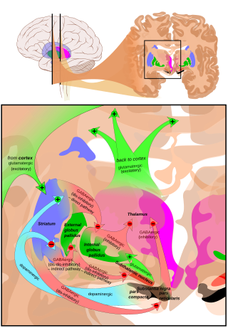
The cortico-basal ganglia-thalamo-cortical loop is a system of neural circuits in the brain. The loop involves connections between the cortex, the basal ganglia, the thalamus, and back to the cortex. It is of particular relevance to hyperkinetic and hypokinetic movement disorders, such as Parkinson's disease and Huntington's disease, as well as to mental disorders of control, such as attention deficit hyperactivity disorder (ADHD), obsessive–compulsive disorder (OCD), and Tourette syndrome.

References

  1. Nauta WJ, Mehler WR (January 1966). "Projections of the lentiform nucleus in the monkey". Brain Res. 1 (1): 3–42. doi:10.1016/0006-8993(66)90103-X. PMID   4956247.
  2. Percheron G, François C, Talbi B, Meder JF, Fenelon G, Yelnik J (1993). "The primate motor thalamus analysed with reference to subcortical afferent territories". Stereotact Funct Neurosurg. 60 (1–3): 32–41. doi:10.1159/000100588. PMID   8511432.
  3. Yelnik J, François C, Percheron G, Tandé D (April 1996). "A spatial and quantitative study of the striatopallidal connection in the monkey". NeuroReport. 7 (5): 985–988. doi:10.1097/00001756-199604100-00006. PMID   8804036. S2CID   25425297.
  4. Schroll, Henning; Hamker, Fred H. (2013-01-01). "Computational models of basal-ganglia pathway functions: focus on functional neuroanatomy". Frontiers in Systems Neuroscience. 7: 122. doi: 10.3389/fnsys.2013.00122 . PMC   3874581 . PMID   24416002.
  5. Morita, Makiko; Hikida, Takatoshi (2015-11-01). "[Distinct roles of the direct and indirect pathways in the basal ganglia circuit mechanism]". Nihon Shinkei Seishin Yakurigaku Zasshi = Japanese Journal of Psychopharmacology. 35 (5–6): 107–111. ISSN   1340-2544. PMID   26785520.
  6. Parent, André; Hazrati, Lili-Naz (1995-01-01). "Functional anatomy of the basal ganglia. II. The place of subthalamic nucleus and external pallidium in basal ganglia circuitry". Brain Research Reviews. 20 (1): 128–154. doi:10.1016/0165-0173(94)00008-D. PMID   7711765. S2CID   20808851.
  7. 1 2 Andrade, Pablo; Carrillo-Ruiz, José D.; Jiménez, Fiacro (2009-07-01). "A systematic review of the efficacy of globus pallidus stimulation in the treatment of Parkinson's disease". Journal of Clinical Neuroscience. 16 (7): 877–881. doi:10.1016/j.jocn.2008.11.006. ISSN   0967-5868. PMID   19398341. S2CID   36080071.
  8. 1 2 Dong, S.; Zhuang, P.; Zhang, X.-H.; Li, J.-Y.; Li, Y.-J. (2012-01-01). "Unilateral deep brain stimulation of the right globus pallidus internus in patients with Tourette's syndrome: two cases with outcomes after 1 year and a brief review of the literature". The Journal of International Medical Research. 40 (5): 2021–2028. doi: 10.1177/030006051204000545 . ISSN   1473-2300. PMID   23206487.
  9. 1 2 Spindler, Meredith A.; Galifianakis, Nicholas B.; Wilkinson, Jayne R.; Duda, John E. (2013-02-01). "Globus pallidus interna deep brain stimulation for tardive dyskinesia: case report and review of the literature". Parkinsonism & Related Disorders. 19 (2): 141–147. doi: 10.1016/j.parkreldis.2012.09.016 . ISSN   1873-5126. PMID   23099106.
  10. Waldvogel, Henry J.; Kim, Eric H.; Tippett, Lynette J.; Vonsattel, Jean-Paul G.; Faull, Richard LM (2014-01-01). Nguyen, Hoa Huu Phuc; Cenci, M. Angela (eds.). Behavioral Neurobiology of Huntington's Disease and Parkinson's Disease. Current Topics in Behavioral Neurosciences. Vol. 22. Springer Berlin Heidelberg. pp. 33–80. doi:10.1007/7854_2014_354. ISBN   9783662463437. PMID   25300927.
  11. Walker, FO (20 January 2007). "Huntington's disease". Lancet. 369 (9557): 218–28. doi:10.1016/S0140-6736(07)60111-1. PMID   17240289. S2CID   46151626.