
In neuroscience and psychology, the term language center refers collectively to the areas of the brain which serve a particular function for speech processing and production. Language is a core system, which gives humans the capacity to solve difficult problems and provides them with a unique type of social interaction. Language allows individuals to attribute symbols to specific concepts and display them through sentences and phrases that follow proper grammatical rules. Moreover, speech is the mechanism in which language is orally expressed.

The brainstem is the posterior stalk-like part of the brain that connects the cerebrum with the spinal cord. In the human brain the brainstem is composed of the midbrain, the pons, and the medulla oblongata. The midbrain is continuous with the thalamus of the diencephalon through the tentorial notch, and sometimes the diencephalon is included in the brainstem.

The corpus callosum, also callosal commissure, is a wide, thick nerve tract, consisting of a flat bundle of commissural fibers, beneath the cerebral cortex in the brain. The corpus callosum is only found in placental mammals. It spans part of the longitudinal fissure, connecting the left and right cerebral hemispheres, enabling communication between them. It is the largest white matter structure in the human brain, about ten centimetres in length and consisting of 200–300 million axonal projections.

Wernicke–Korsakoff syndrome (WKS) is the combined presence of Wernicke encephalopathy (WE) and Korsakoff syndrome. Due to the close relationship between these two disorders, people with either are usually diagnosed with WKS as a single syndrome. It mainly causes vision changes, ataxia and impaired memory.

Wernicke's area, also called Wernicke's speech area, is one of the two parts of the cerebral cortex that are linked to speech, the other being Broca's area. It is involved in the comprehension of written and spoken language, in contrast to Broca's area, which is involved in the production of language. It is traditionally thought to reside in Brodmann area 22, which is located in the superior temporal gyrus in the dominant cerebral hemisphere, which is the left hemisphere in about 95% of right-handed individuals and 70% of left-handed individuals.
Conduction aphasia, also called associative aphasia, is an uncommon form of difficulty in speaking (aphasia). It is caused by damage to the parietal lobe of the brain. An acquired language disorder, it is characterised by intact auditory comprehension, coherent speech production, but poor speech repetition. Affected people are fully capable of understanding what they are hearing, but fail to encode phonological information for production. This deficit is load-sensitive as the person shows significant difficulty repeating phrases, particularly as the phrases increase in length and complexity and as they stumble over words they are attempting to pronounce. People have frequent errors during spontaneous speech, such as substituting or transposing sounds. They are also aware of their errors and will show significant difficulty correcting them.

CarlWernicke was a German physician, anatomist, psychiatrist and neuropathologist. He is known for his influential research into the pathological effects of specific forms of encephalopathy and also the study of receptive aphasia, both of which are commonly associated with Wernicke's name and referred to as Wernicke encephalopathy and Wernicke's aphasia, respectively. His research, along with that of Paul Broca, led to groundbreaking realizations of the localization of brain function, specifically in speech. As such, Wernicke's area has been named after the scientist.

The longitudinal fissure is the deep groove that separates the two cerebral hemispheres of the vertebrate brain. Lying within it is a continuation of the dura mater called the falx cerebri. The inner surfaces of the two hemispheres are convoluted by gyri and sulci just as is the outer surface of the brain.

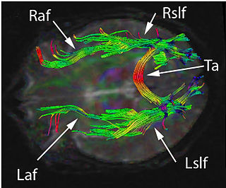
The arcuate fasciculus (AF) is a bundle of axons that generally connects the Broca's area and the Wernicke's area in the brain. It is an association fiber tract connecting caudal temporal cortex and inferior frontal lobe. Fasciculus arcuatus is latin for curved bundle.

The inferior colliculus (IC) is the principal midbrain nucleus of the auditory pathway and receives input from several peripheral brainstem nuclei in the auditory pathway, as well as inputs from the auditory cortex. The inferior colliculus has three subdivisions: the central nucleus, a dorsal cortex by which it is surrounded, and an external cortex which is located laterally. Its bimodal neurons are implicated in auditory-somatosensory interaction, receiving projections from somatosensory nuclei. This multisensory integration may underlie a filtering of self-effected sounds from vocalization, chewing, or respiration activities.

Language processing refers to the way humans use words to communicate ideas and feelings, and how such communications are processed and understood. Language processing is considered to be a uniquely human ability that is not produced with the same grammatical understanding or systematicity in even human's closest primate relatives.
The two-streams hypothesis is a model of the neural processing of vision as well as hearing. The hypothesis, given its initial characterisation in a paper by David Milner and Melvyn A. Goodale in 1992, argues that humans possess two distinct visual systems. Recently there seems to be evidence of two distinct auditory systems as well. As visual information exits the occipital lobe, and as sound leaves the phonological network, it follows two main pathways, or "streams". The ventral stream leads to the temporal lobe, which is involved with object and visual identification and recognition. The dorsal stream leads to the parietal lobe, which is involved with processing the object's spatial location relative to the viewer and with speech repetition.

The anterior commissure is a white matter tract connecting the two temporal lobes of the cerebral hemispheres across the midline, and placed in front of the columns of the fornix. In most existing mammals, the great majority of fibers connecting the two hemispheres travel through the corpus callosum, which is over 10 times larger than the anterior commissure, and other routes of communication pass through the hippocampal commissure or, indirectly, via subcortical connections. Nevertheless, the anterior commissure is a significant pathway that can be clearly distinguished in the brains of all mammals.
The mammillothalamic tract arises from cells in both the medial and lateral nuclei of the mammillary body and by fibers that are directly continued from the fornix.

The inferior longitudinal fasciculus (ILF) is traditionally considered one of the major occipitotemporal association tracts. It is the white matter backbone of the ventral visual stream. It connects the ventral surface of the anterior temporal lobe and the extrastriate cortex of the occipital lobe, running along the lateral and inferior wall of the lateral ventricle.
Amperometry in chemistry is detection of ions in a solution based on electric current or changes in electric current.
Disconnection syndrome is a general term for a collection of neurological symptoms caused -- via lesions to associational or commissural nerve fibres -- by damage to the white matter axons of communication pathways in the cerebrum, independent of any lesions to the cortex. The behavioral effects of such disconnections are relatively predictable in adults. Disconnection syndromes usually reflect circumstances where regions A and B still have their functional specializations except in domains that depend on the interconnections between the two regions.
The mid-fusiform sulcus is a shallow sulcus that divides the fusiform gyrus into lateral and medial partitions. Functionally, the MFS divides both large-scale functional maps and identifies fine-scale functional regions such as the anterior portion of the fusiform face area.
Heinrich Sachs was a late 19th and early 20th century German neurologist and neuroanatomist best known for his atlas of the brain's white matter.
