
The vertebrate cerebrum (brain) is formed by two cerebral hemispheres that are separated by a groove, the longitudinal fissure. The brain can thus be described as being divided into left and right cerebral hemispheres. Each of these hemispheres has an outer layer of grey matter, the cerebral cortex, that is supported by an inner layer of white matter. In eutherian (placental) mammals, the hemispheres are linked by the corpus callosum, a very large bundle of nerve fibers. Smaller commissures, including the anterior commissure, the posterior commissure and the fornix, also join the hemispheres and these are also present in other vertebrates. These commissures transfer information between the two hemispheres to coordinate localized functions.

The occipital lobe is one of the four major lobes of the cerebral cortex in the brain of mammals. The name derives from its position at the back of the head, from the Latin ob, "behind," and caput, "the head."
The cerebrum, telencephalon or endbrain, is the largest part of the brain containing the cerebral cortex, as well as several subcortical structures, including the hippocampus, basal ganglia, and olfactory bulb. In the human brain, the cerebrum is the uppermost region of the central nervous system. The cerebrum develops prenatally from the forebrain (prosencephalon). In mammals, the dorsal telencephalon, or pallium, develops into the cerebral cortex, and the ventral telencephalon, or subpallium, becomes the basal ganglia. The cerebrum is also divided into approximately symmetric left and right cerebral hemispheres.

Wernicke's area, also called Wernicke's speech area, is one of the two parts of the cerebral cortex that are linked to speech, the other being Broca's area. It is involved in the comprehension of written and spoken language, in contrast to Broca's area, which is involved in the production of language. It is traditionally thought to reside in Brodmann area 22, which is located in the superior temporal gyrus in the dominant cerebral hemisphere, which is the left hemisphere in about 95% of right-handed individuals and 70% of left-handed individuals.
Conduction aphasia, also called associative aphasia, is an uncommon form of difficulty in speaking (aphasia). It is caused by damage to the parietal lobe of the brain. An acquired language disorder, it is characterised by intact auditory comprehension, coherent speech production, but poor speech repetition. Affected people are fully capable of understanding what they are hearing, but fail to encode phonological information for production. This deficit is load-sensitive as the person shows significant difficulty repeating phrases, particularly as the phrases increase in length and complexity and as they stumble over words they are attempting to pronounce. People have frequent errors during spontaneous speech, such as substituting or transposing sounds. They are also aware of their errors and will show significant difficulty correcting them.

CarlWernicke was a German physician, anatomist, psychiatrist and neuropathologist. He is known for his influential research into the pathological effects of specific forms of encephalopathy and also the study of receptive aphasia, both of which are commonly associated with Wernicke's name and referred to as Wernicke encephalopathy and Wernicke's aphasia, respectively. His research, along with that of Paul Broca, led to groundbreaking realizations of the localization of brain function, specifically in speech. As such, Wernicke's area has been named after the scientist.

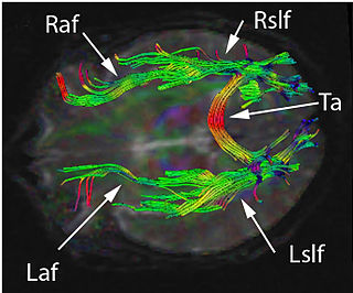
The arcuate fasciculus (AF) is a bundle of axons that generally connects the Broca's area and the Wernicke's area in the brain. It is an association fiber tract connecting caudal temporal cortex and inferior frontal lobe. Fasciculus arcuatus is latin for curved bundle.

Brodmann area 22 is a Brodmann's area that is cytoarchitecturally located in the posterior superior temporal gyrus of the brain. In the left cerebral hemisphere, it is one portion of Wernicke's area. The left hemisphere BA22 helps with generation and understanding of individual words. On the right side of the brain, BA22 helps to discriminate pitch and sound intensity, both of which are necessary to perceive melody and prosody. Wernicke's area is active in processing language and consists of the left Brodmann area 22 and Brodmann area 40, the supramarginal gyrus.

The superior parietal lobule is bounded in front by the upper part of the postcentral sulcus, but is usually connected with the postcentral gyrus above the end of the sulcus. The superior parietal lobule contains Brodmann's areas 5 and 7.

The uncinate fasciculus is a white matter association tract in the human brain that connects parts of the limbic system such as the temporal pole, anterior parahippocampus, and amygdala in the temporal lobe with inferior portions of the frontal lobe such as the orbitofrontal cortex. Its function is unknown though it is affected in several psychiatric conditions. It is one of the last white matter tracts to mature in the human brain.
Constantin von Monakow was a Russian-Swiss neuropathologist who was a native of Bobretsovo in the Vologda Governorate.

The superior longitudinal fasciculus (SLF) is an association tract in the brain that is composed of three separate components. It is present in both hemispheres and can be found lateral to the centrum semiovale and connects the frontal, occipital, parietal, and temporal lobes. This bundle of tracts (fasciculus) passes from the frontal lobe through the operculum to the posterior end of the lateral sulcus where they either radiate to and synapse on neurons in the occipital lobe, or turn downward and forward around the putamen and then radiate to and synapse on neurons in anterior portions of the temporal lobe.

The inferior longitudinal fasciculus (ILF) is traditionally considered one of the major occipitotemporal association tracts. It is the white matter backbone of the ventral visual stream. It connects the ventral surface of the anterior temporal lobe and the extrastriate cortex of the occipital lobe, running along the lateral and inferior wall of the lateral ventricle.
Paul Bucy was an American neurosurgeon and neuropathologist who was a native of Hubbard, Iowa. He is known both for his part in describing the Klüver–Bucy syndrome, his academic life as a teacher in the neurosciences, and for his founding in 1972 and editing Surgical Neurology – An International Journal of Neurosurgery and Neuroscience" from 1972 to 1987.

In human neuroanatomy, brain asymmetry can refer to at least two quite distinct findings:

A nerve tract is a bundle of nerve fibers (axons) connecting nuclei of the central nervous system. In the peripheral nervous system this is known as a nerve, and has associated connective tissue. The main nerve tracts in the central nervous system are of three types: association fibers, commissural fibers, and projection fibers. A tract may also be referred to as a commissure, decussation, pathway or fasciculus. A commissure connects the two cerebral hemispheres at the same levels, while a decussation connects at different levels.
Disconnection syndrome is a general term for a collection of neurological symptoms caused – via lesions to associational or commissural nerve fibres – by damage to the white matter axons of communication pathways in the cerebrum, independent of any lesions to the cortex. The behavioral effects of such disconnections are relatively predictable in adults. Disconnection syndromes usually reflect circumstances where regions A and B still have their functional specializations except in domains that depend on the interconnections between the two regions.
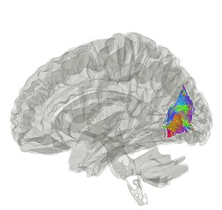
The vertical occipital fasciculus is a fascicle of white matter running vertically in the rear of the brain. It is found at least in primates. It "is the only major fiber bundle connecting dorsolateral and ventrolateral visual cortex."
Lessie Sachs (1897–1942) was a German-born American poet and artist who was active during World War I and World War II.
