Sense of balance

Last updated
Balance skill development in children Balance.JPG
Balance skill development in children
Balance training Plank on two medicine balls.JPG
Balance training

The sense of balance or equilibrioception is the perception of balance and spatial orientation. [1] It helps prevent humans and nonhuman animals from falling over when standing or moving. Equilibrioception is the result of a number of sensory systems working together; the eyes (visual system), the inner ears (vestibular system), and the body's sense of where it is in space (proprioception) ideally need to be intact. [1]

Contents

The vestibular system, the region of the inner ear where three semicircular canals converge, works with the visual system to keep objects in focus when the head is moving. This is called the vestibulo-ocular reflex (VOR). The balance system works with the visual and skeletal systems (the muscles and joints and their sensors) to maintain orientation or balance. Visual signals sent to the brain about the body's position in relation to its surroundings are processed by the brain and compared to information from the vestibular and skeletal systems.

Vestibular system

Diagram of vestibular system Balance Disorder Illustration A.png
Diagram of vestibular system

In the vestibular system, equilibrioception is determined by the level of a fluid called endolymph in the labyrinth, a complex set of tubing in the inner ear.

Dysfunction

This figure shows nerve activity associated with rotational-induced physiologic nystagmus and spontaneous nystagmus resulting from a lesion of one labyrinth. Thin straight arrows show direction of slow components, thick straight arrows show direction of fast components, and curved arrows show direction of endolymph flow in the horizontal semicircular canals. The three semicircular canals are marked AC (anterior canal), PC (posterior canal), and HC (horizontal canal). Balance Disorder Illustration C.png
This figure shows nerve activity associated with rotational-induced physiologic nystagmus and spontaneous nystagmus resulting from a lesion of one labyrinth. Thin straight arrows show direction of slow components, thick straight arrows show direction of fast components, and curved arrows show direction of endolymph flow in the horizontal semicircular canals. The three semicircular canals are marked AC (anterior canal), PC (posterior canal), and HC (horizontal canal).

When the sense of balance is interrupted it causes dizziness, disorientation and nausea. Balance can be upset by Ménière's disease, superior canal dehiscence syndrome, an inner ear infection, by a bad common cold affecting the head or a number of other medical conditions including but not limited to vertigo. It can also be temporarily disturbed by quick or prolonged acceleration, for example, riding on a merry-go-round. Blows can also affect equilibrioreception, especially those to the side of the head or directly to the ear.

Most astronauts find that their sense of balance is impaired when in orbit because they are in a constant state of weightlessness. This causes a form of motion sickness called space adaptation syndrome.

System overview

This diagram linearly (unless otherwise mentioned) tracks the projections of all known structures that allow for balance and acceleration to their relevant endpoints in the human brain. Comprehensive List of Relevant Pathways for the Balance & Acceleration Systems.png
This diagram linearly (unless otherwise mentioned) tracks the projections of all known structures that allow for balance and acceleration to their relevant endpoints in the human brain.
Another diagram showing neural pathway of vestibular/balance system. Arrows show the direction of information relay. Vestibular balance system.jpg
Another diagram showing neural pathway of vestibular/balance system. Arrows show the direction of information relay.

This overview also explains acceleration as its processes are interconnected with balance.

Mechanical

There are five sensory organs innervated by the vestibular nerve; three semicircular canals (Horizontal SCC, Superior SCC, Posterior SCC) and two otolith organs (saccule and utricle). Each semicircular canal (SSC) is a thin tube that doubles in thickness briefly at a point called osseous ampullae. At their center-base, each contains an ampullary cupula. The cupula is a gelatin bulb connected to the stereocilia of hair cells, affected by the relative movement of the endolymph it is bathed in.

Since the cupula is part of the bony labyrinth, it rotates along with actual head movement, and by itself without the endolymph, it cannot be stimulated and therefore, could not detect movement. Endolymph follows the rotation of the canal; however, due to inertia its movement initially lags behind that of the bony labyrinth. The delayed movement of the endolymph bends and activates the cupula. When the cupula bends, the connected stereocilia bend along with it, activating chemical reactions in the hair cells surrounding crista ampullaris and eventually create action potentials carried by the vestibular nerve signaling to the body that it has moved in space.

After any extended rotation, the endolymph catches up to the canal and the cupula returns to its upright position and resets. When extended rotation ceases, however, endolymph continues, (due to inertia) which bends and activates the cupula once again to signal a change in movement. [2]

Pilots doing long banked turns begin to feel upright (no longer turning) as endolymph matches canal rotation; once the pilot exits the turn the cupula is once again stimulated, causing the feeling of turning the other way, rather than flying straight and level.

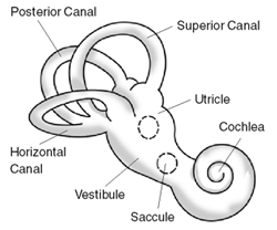
The horizontal SCC handles head rotations about a vertical axis (e.g. looking side to side), the superior SCC handles head movement about a lateral axis (e.g. head to shoulder), and the posterior SCC handles head rotation about a rostral-caudal axis (e.g. nodding). SCC sends adaptive signals, unlike the two otolith organs, the saccule and utricle, whose signals do not adapt over time.[ citation needed ]

A shift in the otolithic membrane that stimulates the cilia is considered the state of the body until the cilia are once again stimulated. For example, lying down stimulates cilia and standing up stimulates cilia, however, for the time spent lying the signal that you are lying remains active, even though the membrane resets.

Otolithic organs have a thick, heavy gelatin membrane that, due to inertia (like endolymph), lags behind and continues ahead past the macula it overlays, bending and activating the contained cilia.

Utricle responds to linear accelerations and head-tilts in the horizontal plane (head to shoulder), whereas saccule responds to linear accelerations and head-tilts in the vertical plane (up and down). Otolithic organs update the brain on the head-location when not moving; SCC update during movement. [3] [4] [5] [6]

Kinocilium are the longest stereocilia and are positioned (one per 40-70 regular cilia) at the end of the bundle. If stereocilia go towards kinocilium, depolarization occurs, causing more neurotransmitters, and more vestibular nerve firings, as compared to when stereocilia tilt away from kinocilium (hyperpolarization, less neurotransmitter, less firing). [7] [8]

Neural

First order vestibular nuclei (VN) project to lateral vestibular nucleus (IVN), medial vestibular nucleus (MVN), and superior vestibular nucleus (SVN).[ clarification needed ]

The inferior cerebellar peduncle is the largest center through which balance information passes. It is the area of integration between proprioceptive, and vestibular inputs, to aid in unconscious maintenance of balance and posture.

The inferior olivary nucleus aids in complex motor tasks by encoding coordinating timing sensory information; this is decoded and acted upon in the cerebellum. [9]

The cerebellar vermis has three main parts. The vestibulocerebellum regulates eye movements by the integration of visual info provided by the superior colliculus and balance information. The spinocerebellum integrates visual, auditory, proprioceptive, and balance information to act out body and limb movements. It receives input from the trigeminal nerve, dorsal column (of the spinal cord), midbrain, thalamus, reticular formation and vestibular nuclei (medulla) outputs[ clarification needed ]. Lastly, the cerebrocerebellum plans, times, and initiates movement after evaluating sensory input from, primarily, motor cortex areas, via pons and cerebellar dentate nucleus. It outputs to the thalamus, motor cortex areas, and red nucleus. [10] [11] [12]

The flocculonodular lobe is a cerebellar lobe that helps maintain body equilibrium by modifying muscle tone (the continuous and passive muscle contractions).

MVN and IVN are in the medulla, LVN and SVN are smaller and in pons. SVN, MVN, and IVN ascend within the medial longitudinal fasciculus. LVN descend the spinal cord within the lateral vestibulospinal tract and ends at the sacrum. MVN also descend the spinal cord, within the medial vestibulospinal tract, ending at lumbar 1. [13] [14]

The thalamic reticular nucleus distributes information to various other thalamic nuclei, regulating the flow of information. It is speculatively able to stop signals, ending transmission of unimportant info. The thalamus relays info between pons (cerebellum link), motor cortices, and insula.

The insula is also heavily connected to motor cortices; the insula is likely where balance is likely brought into perception.

The oculomotor nuclear complex refers to fibers going to tegmentum (eye movement), red nucleus (gait (natural limb movement)), substantia nigra (reward), and cerebral peduncle (motor relay). Nucleus of Cajal are one of the named oculomotor nuclei, they are involved in eye movements and reflex gaze coordination. [15] [16]

The abducens nerve solely innervates the lateral rectus muscle of the eye, moving the eye with the trochlear nerve. The trochlear solely innervates the superior oblique muscle of the eye. Together, trochlear and abducens contract and relax to simultaneously direct the pupil towards an angle and depress the globe on the opposite side of the eye (e.g. looking down directs the pupil down and depresses (towards the brain) the top of the globe). The pupil is not only directed, but often rotated, by these muscles. (See visual system)

The thalamus and superior colliculus are connected via the lateral geniculate nucleus. The superior colliculus (SC) is the topographical map for balance and quick orienting movements with primarily visual inputs. SC integrates multiple senses. [17] [18]

Illustration of the flow of fluid in the ear, which in turn causes displacement of the top portion of the hair cells that are embedded in the jelly-like cupula. Also shows the utricle and saccule organs that are responsible for detecting linear acceleration, or movement in a straight line. Balance Disorder Illustration B.png
Illustration of the flow of fluid in the ear, which in turn causes displacement of the top portion of the hair cells that are embedded in the jelly-like cupula. Also shows the utricle and saccule organs that are responsible for detecting linear acceleration, or movement in a straight line.

Other animals

Some animals have better equilibrioception than humans; for example, a cat uses its inner ear and tail to walk on a thin fence. [19]

Equilibrioception in many marine animals is done with an entirely different organ, the statocyst, which detects the position of tiny calcareous stones to determine which way is "up".

In plants

Plants could be said to exhibit a form of equilibrioception, in that when rotated from their normal attitude the stems grow in the direction that is upward (away from gravity) while their roots grow downward (in the direction of gravity). This phenomenon is known as gravitropism and it has been shown that, for example, poplar stems can detect reorientation and inclination. [20]

Related Research Articles

<span class="mw-page-title-main">Inner ear</span> Innermost part of the vertebrate ear

The inner ear is the innermost part of the vertebrate ear. In vertebrates, the inner ear is mainly responsible for sound detection and balance. In mammals, it consists of the bony labyrinth, a hollow cavity in the temporal bone of the skull with a system of passages comprising two main functional parts:

Articles related to anatomy include:

<span class="mw-page-title-main">Brainstem</span> Posterior part of the brain, adjoining and structurally continuous

The brainstem is the posterior stalk-like part of the brain that connects the cerebrum with the spinal cord. In the human brain the brainstem is composed of the midbrain, the pons, and the medulla oblongata. The midbrain is continuous with the thalamus of the diencephalon through the tentorial notch, and sometimes the diencephalon is included in the brainstem.

<span class="mw-page-title-main">Semicircular canals</span> Organ located in innermost part of ear

The semicircular canals or semicircular ducts are three semicircular, interconnected tubes located in the innermost part of each ear, the inner ear. The three canals are the horizontal, superior and posterior semicircular canals. [This section needs editing - different terms are used in diagram and later in the text.]

<span class="mw-page-title-main">Vestibulo–ocular reflex</span>

The vestibulo-ocular reflex (VOR) is a reflex acting to stabilize gaze during head movement, with eye movement due to activation of the vestibular system. The reflex acts to stabilize images on the retinas of the eye during head movement. Gaze is held steadily on a location by producing eye movements in the direction opposite that of head movement. For example, when the head moves to the right, the eyes move to the left, meaning the image a person sees stays the same even though the head has turned. Since slight head movement is present all the time, VOR is necessary for stabilizing vision: people with an impaired reflex find it difficult to read using print, because the eyes do not stabilise during small head tremors, and also because damage to reflex can cause nystagmus.

<span class="mw-page-title-main">Utricle (ear)</span> Membranous labyrinth in the vestibule of ear

The utricle and saccule are the two otolith organs in the vertebrate inner ear. They are part of the balancing system in the vestibule of the bony labyrinth. They use small stones and a viscous fluid to stimulate hair cells to detect motion and orientation. The utricle detects linear accelerations and head-tilts in the horizontal plane. The word utricle comes from Latin uter 'leather bag'.

<span class="mw-page-title-main">Saccule</span> Bed of sensory cells in the inner ear

The saccule is a bed of sensory cells in the inner ear. It translates head movements into neural impulses for the brain to interpret. The saccule detects linear accelerations and head tilts in the vertical plane. When the head moves vertically, the sensory cells of the saccule are disturbed and the neurons connected to them begin transmitting impulses to the brain. These impulses travel along the vestibular portion of the eighth cranial nerve to the vestibular nuclei in the brainstem.

<span class="mw-page-title-main">Vestibular system</span> Sensory system that facilitates body balance

The vestibular system, in vertebrates, is a sensory system that creates the sense of balance and spatial orientation for the purpose of coordinating movement with balance. Together with the cochlea, a part of the auditory system, it constitutes the labyrinth of the inner ear in most mammals.

<span class="mw-page-title-main">Auditory system</span> Sensory system used for hearing

The auditory system is the sensory system for the sense of hearing. It includes both the sensory organs and the auditory parts of the sensory system.

<span class="mw-page-title-main">Endolymph</span> Inner ear fluid

Endolymph is the fluid contained in the membranous labyrinth of the inner ear. The major cation in endolymph is potassium, with the values of sodium and potassium concentration in the endolymph being 0.91 mM and 154 mM, respectively. It is also called Scarpa's fluid, after Antonio Scarpa.

<span class="mw-page-title-main">Pontine tegmentum</span>

The pontine tegmentum, or dorsal pons, is located within the brainstem, and is one of two parts of the pons, the other being the ventral pons or basilar part of the pons. The pontine tegmentum can be defined in contrast to the basilar pons: basilar pons contains the corticospinal tract running craniocaudally and can be considered the rostral extension of the ventral medulla oblongata; however, basilar pons is distinguished from ventral medulla oblongata in that it contains additional transverse pontine fibres that continue laterally to become the middle cerebellar peduncle. The pontine tegmentum is all the material dorsal from the basilar pons to the fourth ventricle. Along with the dorsal surface of the medulla, it forms part of the rhomboid fossa – the floor of the fourth ventricle.

<span class="mw-page-title-main">Stereocilia (inner ear)</span>

In the inner ear, stereocilia are the mechanosensing organelles of hair cells, which respond to fluid motion in numerous types of animals for various functions, including hearing and balance. They are about 10–50 micrometers in length and share some similar features of microvilli. The hair cells turn the fluid pressure and other mechanical stimuli into electric stimuli via the many microvilli that make up stereocilia rods. Stereocilia exist in the auditory and vestibular systems.

<span class="mw-page-title-main">Flocculus</span>

The flocculus is a small lobe of the cerebellum at the posterior border of the middle cerebellar peduncle anterior to the biventer lobule. Like other parts of the cerebellum, the flocculus is involved in motor control. It is an essential part of the vestibulo-ocular reflex, and aids in the learning of basic motor skills in the brain.

<span class="mw-page-title-main">Vestibulospinal tract</span>

The vestibulospinal tract is a neural tract in the central nervous system. Specifically, it is a component of the extrapyramidal system and is classified as a component of the medial pathway. Like other descending motor pathways, the vestibulospinal fibers of the tract relay information from nuclei to motor neurons. The vestibular nuclei receive information through the vestibulocochlear nerve about changes in the orientation of the head. The nuclei relay motor commands through the vestibulospinal tract. The function of these motor commands is to alter muscle tone, extend, and change the position of the limbs and head with the goal of supporting posture and maintaining balance of the body and head.

<span class="mw-page-title-main">Ampullary cupula</span>

The ampullary cupula, or cupula, is a structure in the vestibular system, providing the sense of spatial orientation.

<span class="mw-page-title-main">Otolithic membrane</span>

The otolithic membrane is a fibrous structure located in the vestibular system of the inner ear. It plays a critical role in the brain's interpretation of equilibrium. The membrane serves to determine if the body or the head is tilted, in addition to the linear acceleration of the body. The linear acceleration could be in the horizontal direction as in a moving car or vertical acceleration such as that felt when an elevator moves up or down.

A kinocilium is a special type of cilium on the apex of hair cells located in the sensory epithelium of the vertebrate inner ear.

<span class="mw-page-title-main">Crista ampullaris</span>

The crista ampullaris is the sensory organ of rotation. They are found in the ampullae of each of the semicircular canals of the inner ear, meaning that there are three pairs in total. The function of the crista ampullaris is to sense angular acceleration and deceleration.

The vestibular evoked myogenic potential is a neurophysiological assessment technique used to determine the function of the otolithic organs of the inner ear. It complements the information provided by caloric testing and other forms of inner ear testing. There are two different types of VEMPs. One is the oVEMP and another is the cVEMP. The oVEMP measures integrity of the utricule and superior vestibular nerve and the cVemp measures the saccule and the inferior vestibular nerve.

The righting reflex, also known as the labyrinthine righting reflex, is a reflex that corrects the orientation of the body when it is taken out of its normal upright position. It is initiated by the vestibular system, which detects that the body is not erect and causes the head to move back into position as the rest of the body follows. The perception of head movement involves the body sensing linear acceleration or the force of gravity through the otoliths, and angular acceleration through the semicircular canals. The reflex uses a combination of visual system inputs, vestibular inputs, and somatosensory inputs to make postural adjustments when the body becomes displaced from its normal vertical position. These inputs are used to create what is called an efference copy. This means that the brain makes comparisons in the cerebellum between expected posture and perceived posture, and corrects for the difference. The reflex takes 6 or 7 weeks to perfect, but can be affected by various types of balance disorders.

References

  1. 1 2 Wolfe, Jeremy; Kluender, Keith; Levi, Dennis (2012). Sensation & perception (3rd ed.). Sinauer Associates. p. 7. ISBN   978-0878935727.
  2. Seeley, R., VanPutte, C., Regan, J., & Russo, A. (2011). Seeley's Anatomy & Physiology (9th ed.). New York: McGraw Hill [ ISBN missing ][ page needed ]
  3. Albertine, Kurt. Barron’s Anatomy Flash Cards
  4. "How Does Our Sense of Balance Work?" How Does Our Sense of Balance Work?U.S. National Library of Medicine, 12 Jan. 2012.
  5. "Semicircular Canals." Semicircular Canals Function, Definition & Anatomy. Healthline Medical Team, 26 Jan. 2015.
  6. Tillotson, Joanne. McCann, Stephanie. Kaplan’s Medical Flashcards. Apr. 02. 2013.
  7. Spoor, Fred, and Theodore Garland, Jr. "The Primate Semicircular Canal System and Locomotion." 8 May 2007.
  8. Sobkowicz, H.M., and S.M. Slapnick. "The Kinocilium of Auditory Hair Cells and Evidence for Its Morphogenet." Ic Role during the Regeneration of Stereocilia and Cuticular Plates. Sept. 1995.
  9. Mathy, Alexandre, and Sara S.N. Ho. "Encoding of Oscillations by Axonal Bursts in Inferior Olive Neurons." Science Direct. 14 May 2009. Web. 28 Mar. 2016.
  10. Chen, S.H. Annabel, and John E. Desmond. "Cerebrocerebellar Networks during Articulatory Rehearsal and Verbal Working Memory Tasks." Science Direct. 15 Jan. 2005. Web. 28 Mar. 2016.
  11. Barmack, Neil H. "Central Vestibular System: Vestibular Nuclei and Posterior Cerebellum." Science Direct. 15 June 2003. Web. 28 Mar. 2016.
  12. Akiyama, K., and S. Takazawa. "Bilateral Middle Cerebellar Peduncle Infarction Caused by Traumatic Vertebral Artery Dissection." JNeurosci. 1 Mar. 2001. 28 Mar. 2016.
  13. Gdowski, Greg T., and Robert A. McCrea. "Integration of Vestibular and Head Movement Signals in the Vestibular Nuclei During Whole-Body Rotation. 01 July 1999. Web. 28 Mar. 2016.
  14. Roy, Jefferson E., and Kathleen E. Cullen. "Dissociating Self-Generated from Passively Applied Head Motion: Neural Mechanisms in the Vestibular Nuclei." JNeurosci. 3 Mar. 2004. Web. 28 Mar. 2016.
  15. Takagi, Mineo, and David S. Zee. "Effects of Lesions of the Oculomotor Cerebellar Vermis on Eye Movements in Primate: Smooth Pursuit." 01 Apr. 2000
  16. Klier, Eliana M., and Hongying Wang. "Interstitial Nucleus of Cajal Encodes Three-Dimensional Head Orientations in Fick-Like Coordinates." Articles, 01 Jan. 2007.
  17. May, Paul J. "The Mammalian Superior Colliculus: Laminar Structure and Connections." Science Direct. 2006.
  18. Corneil, Brian D., and Etienne Olivier. "Neck Muscle Responses to Stimulation of Monkey Superior Colliculus. I. Topography and Manipulation of Stimulation Parameters." 01 Oct. 2002. Web. 28 Mar. 2016.
  19. "Equilibrioception". ScienceDaily . Archived from the original on 18 May 2011. Retrieved 15 January 2011.
  20. Azri, W.; Chambon, C.; Herbette, S. P.; Brunel, N.; Coutand, C.; Leplé, J. C.; Ben Rejeb, I.; Ammar, S.; Julien, J. L.; Roeckel-Drevet, P. (2009). "Proteome analysis of apical and basal regions of poplar stems under gravitropic stimulation". Physiologia Plantarum . 136 (2): 193–208. doi:10.1111/j.1399-3054.2009.01230.x. PMID   19453506.