Topographic map (neuroanatomy)

Last updated

In neuroanatomy, topographic map is the ordered projection of a sensory surface (like the retina or the skin) or an effector system (like the musculature) to one or more structures of the central nervous system. Topographic maps can be found in all sensory systems and in many motor systems.

Contents

Visual system

The visual system refers to the part of the central nervous system that allows an organism to see. It interprets information from visible light to build a representation of the world. The ganglion cells of the retina project in an orderly fashion to the lateral geniculate nucleus (LGN) of the thalamus and from there to the primary visual cortex (V1); adjacent spots on the retina are represented by adjacent neurons in the lateral geniculate nucleus and the primary visual cortex. The term for this pattern of projection is topography. There are many types of topographic maps in the visual cortices, including retinotopic maps, ocular dominance maps and orientation maps. Retinotopic maps are the easiest to understand in terms of topography. Retinotopic maps are those in which the image on the retina is maintained in the cortices (V1 and the LGN). In other words, if a specific region of the cortices was damaged, that individual would then have a blind spot in the real world, they would not be able to see the bit of the world that corresponded to the retina damage. Orientation maps are also topographic. In these maps there are cells which have a preference to a certain orientation, the maximum firing rate of the cell will be achieved at that preference. As the orientation is moved away from, the firing rate will drop. An orientation map is topographic because neighboring neural tissues have similar orientation preferences.

The term retinotopic refers to the maintenance of the particular order of afferent connections from the retina along the afferent pathway via sub-cortical structures to V1 and other cortical visual areas. The primary visual cortex (V1, Brodmann's area 17) is the first cortical area to receive visual input. The stria of Gennari – a set of heavily myelinated, horizontally projecting axons within the termination zone of lateral geniculate nucleus (LGN) input to V1 – provides an anatomical marker particular to V1. [1]

Development

According to the chemoaffinity hypothesis, chemical labels are distributed in a graded fashion across the retina and tectum. This allows each retinal ganglion cell to recognize its proper termination site. Experiments with artificially created compound eyes in Xenopus demonstrate that not only the ganglion cells but also their axons carry these specificities. Axons must be able to communicate with each other to ensure that ones with the same positional tags innervate the same area of the superior colliculus. [2]

Categories of retinotopic maps

First-order representations are those in which adjacent points of the same hemifield always map to adjacent columns in the contralateral cortex. An example of this would be the map in primary visual cortex (V1).

Second-order representations, also known as a field discontinuity map, are maps that are organized such that it appears that a discontinuity has been introduced in either the visual field or the retina. The maps in V2 and other extrastriate cortex are second-order representations. [3]

Auditory system

The auditory system is the sensory system for hearing in which the brain interprets information from the frequency of sound waves, yielding the perception of tones. Sound waves enter the ear through the auditory canal. These waves arrive at the eardrum where the properties of the waves are transduced into vibrations. The vibrations travel through the bones of the inner ear to the cochlea. In the cochlea, the vibrations are transduced into electrical information through the firing of hair cells in the organ of Corti. The organ of Corti projects in an orderly fashion to structures in the brainstem (namely, the cochlear nuclei and the inferior colliculus), and from there to the medial geniculate nucleus of the thalamus and the primary auditory cortex. Adjacent sites on the organ of Corti, which are themselves selective for the sound frequency, are represented by adjacent neurons in the aforementioned CNS structures. This projection pattern has been termed tonotopy.

The tonotopic layout of sound information begins in the cochlea where the basilar membrane vibrates at different positions along its length depending upon the frequency of the sound. Higher frequency sounds are at the base of the cochlea, if it were unrolled, and low frequency sounds are at the apex. This arrangement is also found in the auditory cortex in the temporal lobe. In areas that are tonotopically organized, the frequency varies systematically from low to high along the surface of the cortex, but is relatively constant across cortical depth. The general image of topographic organization in animals is multiple tonotopic maps distributed over the surface of the cortex. [4]

Somatosensory system

The somatosensory system comprises a diverse range of receptors and processing centers to produce the perception of touch, temperature, proprioception, and nociception. Receptors are located throughout the body including the skin, epithelia, internal organs, skeletal muscles, bones, and joints. The cutaneous receptors of the skin project in an orderly fashion to the spinal cord, and from there, via different afferent pathways (dorsal column-medial lemniscus tract and spinothalamic tract), to the ventral posterior nucleus of the thalamus and the primary somatosensory cortex. Again, adjacent areas on the skin are represented by adjacent neurons in all aforementioned structures. This projection pattern has been termed somatotopy.

One common diagram of the somatotopic map is the cortical homunculus. This illustration is a fairly accurate representation of how much cortical area represents each body part or region. It also maps what part of the cortex represents each region of the body.

Motor system

Unlike the topographic maps of the senses, the neurons of the motor cortex are efferent neurons that exit the brain instead of bringing information to the brain through afferent connections. The motor system is responsible for initiating voluntary or planned movements (reflexes are mediated at the spinal cord level, so movements associated with a reflex are not initiated by the motor cortex). The activation from the motor cortex travels through Betz cells down the corticospinal tract through upper motor neurons, terminating at the anterior horn of the grey matter where lower motor neurons transmit the signal to peripheral motor neurons and, finally, the voluntary muscles.

Movements not requiring use of topographic maps

There are several instances of movements that do not require the participation of topographic maps, or even the participation of the brain. One instance would be (as already stated) in some reflexes. Reflexes usually are mediated at the level of the spinal cord through reflex arcs. In humans, mono-, oligo-, and poly-synaptic reflex arcs, propriospinal interneuron systems, and internuncial gray matter neurons collectively participate continuously to produce spinal cord reflex that activates muscle. [5]

The higher motor centers of octopuses (large brained invertebrates) are notable for organizing (unlike vertebrates) highly skilled movements without the use of somatotopic maps of their bodies. [6]

Olfactory system

The olfactory system is the sensory system used for olfaction, or the sense of smell. It detects volatile, airborne substances.

Most sensory systems spatially segregate afferent input from primary sensory neurons to construct a topographic map that defines the location of a sensory stimulus within the environment, as well as the quality of the stimulus itself. Unlike other sensory systems, the topography in the olfactory system is not dependent on spatial properties of the stimuli. Relieved of the requirement to map the position of an olfactory stimulus in space, the olfactory system employs spatial segregation of sensory input to encode the quality of an odorant. [7]

The topographic map revealed in the olfactory system differs in quality from the orderly representation inherent in the retinotopic, tonotopic, or somatotopic sensory maps. Olfactory neurons differ from one another by the nature of the receptor that they possess. However, the olfactory sensory neurons to which olfactory bulb neurons are connected are also distributed across the receptor sheet (the olfactory epithelium) depending on their chemical preferences. Locations on the olfactory epithelium and the olfactory bulb are correlated [8] so, as with other sensory systems, the topographic map in the brain is linked to the structure of the peripheral organ. This principle is called rhinotopy. [7]

Individual olfactory sensory neurons express only one of the thousand receptor genes, such that neurons are functionally distinct. Cells expressing a given receptor in the olfactory epithelium are randomly dispersed within 1 of 4 broad zones. Sensory neurons extend a single unbranched axon to the olfactory bulb such that the projections from neurons expressing a specific receptor converge on 2 out of the 1800 glomeruli. [9] The pattern of convergence is absolute and invariant in all individuals in a species. The bulb therefore provides a spatial map that identifies which of the numerous receptors have been activated within the sensory epithelium so that the quality of an olfactory stimulus is encoded by specific combination of glomeruli activated by a given odorant (combinatorial code).

While refinement of the bulbar topographic code relies on activity, the development occurs partly without apparent contribution from activity-dependent processes. Mice lacking the olfactory cyclic nucleotide-gated ion channel fail to exhibit odor-evoked electrophysiological responses in the sensory epithelium, but the pattern of convergence of like axons in the bulb is unaltered in these mutant mice, arguing strongly that olfactory experience is not necessary for the establishment or refinement of the topographic map.

These findings, however, do not exclude a role for activity-dependent processes in the maintenance or potential plasticity of the map after it is established. For example, neurons that do not express functional odorant receptors have been observed to have a significantly shorter half-life. Activity-dependent processes may therefore be essential for the survival of the olfactory neurons postnatally, and in that manner, may alter the input to individual glomeruli, altering the sensitivity to individual odors. [10]

Gustatory system

The gustatory system is the sensory system for taste. Like olfaction, taste requires a process of chemoreception. The receptors for taste are taste buds on the tongue. The tongue contains taste receptors, as well as mechanoreceptors. Afferents from taste receptors and mechanoreceptors of the tongue access different ascending systems in the brainstem. However, it is uncertain how these two sources of information are processed in cortex. The primary gustatory cortex (G) is located near the somatotopic region for the tongue (S1), in the insular cortex deep in the lateral fissure with the secondary taste areas in the opercula. [11]

The peripheral taste system likely maintains a specific relationship between taste bud cells selectively responsive to one taste quality and the ganglion cells signaling that particular quality. This explains the response specificity of some individual taste nerve, particularly because sweet, amino acid, and bitter receptors are expressed in distinct populations of taste cells. Although anatomical evidence for such an exclusive relationship is lacking at the level of single receptor and ganglion cells, the relationship between single buds and their innervating ganglion cells is tractable neuroanatomically. In taste, attempts to identify a spatial representation of taste receptors or taste qualities have revealed only an indistinct functional topography in the brain. Nevertheless, taste ganglion cells must distribute peripheral fibers to particular receptor cell types and disseminate impulses centrally in a structurally organized manner. [12]

Benefits

For neurons, it is metabolically costly to make long connections, and the trade-off of connection distance is time. Thus, it is good design to group neurons together that are to be highly interconnected. Multiple topographic maps is a feature that is advantageous because it allows maps of different sizes that would accommodate varying levels of acuity and details in signals. A more detailed map has more neurons that would take up more area than a more global map, which would require fewer connections. [13]

Techniques

A variety of techniques have been used to establish the topographic maps in the brain. The existence of topographical maps was shown early by electrical stimulation of the cortex, tracing patterns of epileptic seizures, stimulation sequences, and impairments due to lesions. Details in the maps came later through microelectrode stimulation and recording techniques became commonly used in demonstrating somatotopic maps and later in the auditory and visual systems, both cortically and in subcortical structures such as the colliculi and geniculate nuclei of the thalamus. [14] Single-cell recording, transcranial magnetic stimulation (TMS), electrical stimulation of the cortex, and functional magnetic resonance imaging (fMRI) are some of the techniques used to study maps in the brain. Many of the existing topographic maps have been further studied or refined using fMRI. For example, Hubel and Wiesel originally studied the retinotopic maps in the primary visual cortex using single-cell recording. Recently, however, imaging of the retinotopic map in the cortex and in sub-cortical areas, such as the lateral geniculate nucleus, have been improved using the fMRI technique. [15]

See also

Related Research Articles

<span class="mw-page-title-main">Visual cortex</span> Region of the brain that processes visual information

The visual cortex of the brain is the area of the cerebral cortex that processes visual information. It is located in the occipital lobe. Sensory input originating from the eyes travels through the lateral geniculate nucleus in the thalamus and then reaches the visual cortex. The area of the visual cortex that receives the sensory input from the lateral geniculate nucleus is the primary visual cortex, also known as visual area 1 (V1), Brodmann area 17, or the striate cortex. The extrastriate areas consist of visual areas 2, 3, 4, and 5.

<span class="mw-page-title-main">Cerebral cortex</span> Outer layer of the cerebrum of the mammalian brain

The cerebral cortex, also known as the cerebral mantle, is the outer layer of neural tissue of the cerebrum of the brain in humans and other mammals. It is the largest site of neural integration in the central nervous system, and plays a key role in attention, perception, awareness, thought, memory, language, and consciousness. The cerebral cortex is the part of the brain responsible for cognition.

<span class="mw-page-title-main">Trigeminal nerve</span> Cranial nerve responsible for the faces senses and motor functions

In neuroanatomy, the trigeminal nerve (lit. triplet nerve), also known as the fifth cranial nerve, cranial nerve V, or simply CN V, is a cranial nerve responsible for sensation in the face and motor functions such as biting and chewing; it is the most complex of the cranial nerves. Its name (trigeminal, from Latin tri- 'three' and -geminus 'twin') derives from each of the two nerves (one on each side of the pons) having three major branches: the ophthalmic nerve (V1), the maxillary nerve (V2), and the mandibular nerve (V3). The ophthalmic and maxillary nerves are purely sensory, whereas the mandibular nerve supplies motor as well as sensory (or "cutaneous") functions. Adding to the complexity of this nerve is that autonomic nerve fibers as well as special sensory fibers (taste) are contained within it.

<span class="mw-page-title-main">Visual system</span> Body parts responsible for vision

The visual system is the physiological basis of visual perception. The system detects, transduces and interprets information concerning light within the visible range to construct an image and build a mental model of the surrounding environment. The visual system is associated with the eye and functionally divided into the optical system and the neural system.

<span class="mw-page-title-main">Sensory nervous system</span> Part of the nervous system

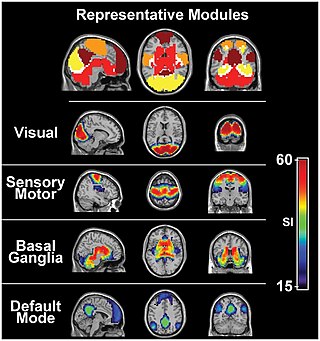
The sensory nervous system is a part of the nervous system responsible for processing sensory information. A sensory system consists of sensory neurons, neural pathways, and parts of the brain involved in sensory perception and interoception. Commonly recognized sensory systems are those for vision, hearing, touch, taste, smell, balance and visceral sensation. Sense organs are transducers that convert data from the outer physical world to the realm of the mind where people interpret the information, creating their perception of the world around them.

<span class="mw-page-title-main">Lateral geniculate nucleus</span> Component of the visual system in the brains thalamus

In neuroanatomy, the lateral geniculate nucleus is a structure in the thalamus and a key component of the mammalian visual pathway. It is a small, ovoid, ventral projection of the thalamus where the thalamus connects with the optic nerve. There are two LGNs, one on the left and another on the right side of the thalamus. In humans, both LGNs have six layers of neurons alternating with optic fibers.

<span class="mw-page-title-main">Brodmann area</span> 52 distinct regions of the brains cerebral cortex

A Brodmann area is a region of the cerebral cortex, in the human or other primate brain, defined by its cytoarchitecture, or histological structure and organization of cells. The concept was first introduced by the German anatomist Korbinian Brodmann in the early 20th century. Brodmann mapped the human brain based on the varied cellular structure across the cortex and identified 52 distinct regions, which he numbered 1 to 52. These regions, or Brodmann areas, correspond with diverse functions including sensation, motor control, and cognition.

<span class="mw-page-title-main">Claustrum</span> Structure in the brain

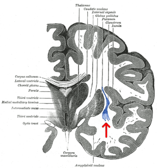
The claustrum is a thin sheet of neurons and supporting glial cells in the brain, that connects to the cerebral cortex and subcortical regions including the amygdala, hippocampus and thalamus. It is located between the insular cortex laterally and the putamen medially, encased by the extreme and external capsules respectively. Blood to the claustrum is supplied by the middle cerebral artery. It is considered to be the most densely connected structure in the brain, and thus hypothesized to allow for the integration of various cortical inputs such as vision, sound and touch, into one experience. Other hypotheses suggest that the claustrum plays a role in salience processing, to direct attention towards the most behaviorally relevant stimuli amongst the background noise. The claustrum is difficult to study given the limited number of individuals with claustral lesions and the poor resolution of neuroimaging.

<span class="mw-page-title-main">Sensory neuron</span> Nerve cell that converts environmental stimuli into corresponding internal stimuli

Sensory neurons, also known as afferent neurons, are neurons in the nervous system, that convert a specific type of stimulus, via their receptors, into action potentials or graded receptor potentials. This process is called sensory transduction. The cell bodies of the sensory neurons are located in the dorsal root ganglia of the spinal cord.

The receptive field, or sensory space, is a delimited medium where some physiological stimuli can evoke a sensory neuronal response in specific organisms.

<span class="mw-page-title-main">Superior colliculus</span> Structure in the midbrain

In neuroanatomy, the superior colliculus is a structure lying on the roof of the mammalian midbrain. In non-mammalian vertebrates, the homologous structure is known as the optic tectum or optic lobe. The adjective form tectal is commonly used for both structures.

<span class="mw-page-title-main">Thalamocortical radiations</span> Neural pathways between the thalamus and cerebral cortex

In neuroanatomy, thalamocortical radiations, also known as thalamocortical fibers, are the efferent fibers that project from the thalamus to distinct areas of the cerebral cortex. They form fiber bundles that emerge from the lateral surface of the thalamus.

<span class="mw-page-title-main">Retinotopy</span> Mapping of visual input from the retina to neurons

Retinotopy is the mapping of visual input from the retina to neurons, particularly those neurons within the visual stream. For clarity, 'retinotopy' can be replaced with 'retinal mapping', and 'retinotopic' with 'retinally mapped'.

In medicine and anatomy, the special senses are the senses that have specialized organs devoted to them:

<span class="mw-page-title-main">Bipolar neuron</span> Neuron with only one axon and one dendrite

A bipolar neuron, or bipolar cell, is a type of neuron characterized by having both an axon and a dendrite extending from the soma in opposite directions. These neurons are predominantly found in the retina and olfactory system. The embryological period encompassing weeks seven through eight marks the commencement of bipolar neuron development.

The isothalamus is a division used by some researchers in describing the thalamus.

A sensory map is an area of the brain which responds to sensory stimulation, and are spatially organized according to some feature of the sensory stimulation. In some cases the sensory map is simply a topographic representation of a sensory surface such as the skin, cochlea, or retina. In other cases it represents other stimulus properties resulting from neuronal computation and is generally ordered in a manner that reflects the periphery. An example is the somatosensory map which is a projection of the skin's surface in the brain that arranges the processing of tactile sensation. This type of somatotopic map is the most common, possibly because it allows for physically neighboring areas of the brain to react to physically similar stimuli in the periphery or because it allows for greater motor control.

Feature detection is a process by which the nervous system sorts or filters complex natural stimuli in order to extract behaviorally relevant cues that have a high probability of being associated with important objects or organisms in their environment, as opposed to irrelevant background or noise.

<span class="mw-page-title-main">Nucleus basalis</span> Group of neurons in the brain

In the human brain, the nucleus basalis, also known as the nucleus basalis of Meynert or nucleus basalis magnocellularis, is a group of neurons located mainly in the substantia innominata of the basal forebrain. Most neurons of the nucleus basalis are rich in the neurotransmitter acetylcholine, and they have widespread projections to the neocortex and other brain structures.

<span class="mw-page-title-main">Michel Imbert</span> French neuropsychologist

Michel Imbert is a neuropsychologist teacher-researcher in cognitive neurosciences, professor emeritus at Pierre-et-Marie-Curie University and honorary director of studies at the École des hautes études en sciences sociales (EHESS).

References

  1. Hinds, O.; et al. (2009). "Locating the functional and anatomical boundaries of human primary visual cortex". NeuroImage. 46 (4): 915–922. doi:10.1016/j.neuroimage.2009.03.036. PMC   2712139 . PMID   19328238.
  2. Udin, S.; Fawcett, J. (1988). "Formation of Topographic Maps". Annu. Rev. Neurosci. 11: 289–327. doi:10.1146/annurev.ne.11.030188.001445. PMID   3284443.
  3. Rosa, M.G.P. (2002). "Visual maps in the adult primate cerebral cortex: some implications for brain development and evolution". Braz J Med Biol Res. 35 (12): 1485–1498. doi: 10.1590/s0100-879x2002001200008 . PMID   12436190.
  4. Talavage, T. M.; et al. (2000). "Frequency-dependent responses exhibited by multiple regions in human auditory cortex". Hearing Research. 150 (1–2): 225–244. doi:10.1016/s0378-5955(00)00203-3. PMID   11077206. S2CID   8678128.
  5. Dimitrijevic, M. R., et al. (2005). Motor Control in the Human Spinal Cord. Artificial Organs. 29(3): 216-219.
  6. Zullo L, Sumbre G, Agnisola C, Flash T, Hochner B. (2009). Nonsomatotopic organization of the higher motor centers in octopus. Curr Biol. 19(19):1632-6.
  7. 1 2 Auffarth, B (2013). "Understanding smell -- the olfactory stimulus problem". Neuroscience & Biobehavioral Reviews. 37 (8): 1667–1679. doi:10.1016/j.neubiorev.2013.06.009. PMID   23806440. S2CID   207090474.
  8. Imai, T.; et al. (2010). "Topographic Mapping—The Olfactory System". Cold Spring Harb Perspect Biol. 2 (8): 8. doi:10.1101/cshperspect.a001776. PMC   2908763 . PMID   20554703.
  9. Vosshall, L.; et al. (2000). "An Olfactory Sensory Map in the Fly Brain". Cell. 102 (2): 147–159. doi: 10.1016/s0092-8674(00)00021-0 . PMID   10943836. S2CID   17573876.
  10. Wang, F; Nemes, A; Meldelson, M; Axel, R (1998). "Odorant Receptors Govern the Formation of a Precise Topographic Map". Cell. 93 (1): 47–60. doi: 10.1016/s0092-8674(00)81145-9 . PMID   9546391. S2CID   18458277.
  11. Kaas, J. H., Qi, H. X., & Iyengar, S. (2006). Cortical network for representing the teeth and tongue in primates. Anatomical Record Part a-Discoveries in Molecular Cellular and Evolutionary Biology, 288A(2): 182-190.
  12. Zaidi, F. N.; Whitehead, M. C. (2006). "Discrete Innervation of Murine Taste Buds by Peripheral Taste Neurons". J. Neurosci. 26 (32): 8243–8253. doi: 10.1523/jneurosci.5142-05.2006 . PMC   6673808 . PMID   16899719.
  13. Kaas, J. H. (2008). "The evolution of the complex sensory and motor systems of the human brain". Brain Research Bulletin. 75 (2–4): 384–390. doi:10.1016/j.brainresbull.2007.10.009. PMC   2349093 . PMID   18331903.
  14. Kaas, J. H. (1997). "Topographic Maps are Fundamental to Sensory Processing". Brain Research Bulletin. 44 (2): 107–112. doi:10.1016/s0361-9230(97)00094-4. PMID   9292198. S2CID   14488006.
  15. Schneider, KA; Richter, MC; Kastner, S (2004). "Retinotopic Organization and Functional Subdivision of the Human Lateral Geniculate Nucleus: A High-Resolution Functional Magnetic Resonance Imaging Study". J. Neurosci. 24 (41): 8975–8985. doi: 10.1523/jneurosci.2413-04.2004 . PMC   6730047 . PMID   15483116.

Further reading