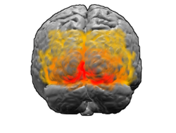
The visual cortex of the brain is the area of the cerebral cortex that processes visual information. It is located in the occipital lobe. Sensory input originating from the eyes travels through the lateral geniculate nucleus in the thalamus and then reaches the visual cortex. The area of the visual cortex that receives the sensory input from the lateral geniculate nucleus is the primary visual cortex, also known as visual area 1 (V1), Brodmann area 17, or the striate cortex. The extrastriate areas consist of visual areas 2, 3, 4, and 5.
Blindsight is the ability of people who are cortically blind to respond to visual stimuli that they do not consciously see due to lesions in the primary visual cortex, also known as the striate cortex or Brodmann Area 17. The term was coined by Lawrence Weiskrantz and his colleagues in a paper published in a 1974 issue of Brain. A previous paper studying the discriminatory capacity of a cortically blind patient was published in Nature in 1973. The assumed existence of blindsight is controversial, with some arguing that it is merely degraded conscious vision.

The visual system is the physiological basis of visual perception. The system detects, transduces and interprets information concerning light within the visible range to construct an image and build a mental model of the surrounding environment. The visual system is associated with the eye and functionally divided into the optical system and the neural system.
The sensory nervous system is a part of the nervous system responsible for processing sensory information. A sensory system consists of sensory neurons, neural pathways, and parts of the brain involved in sensory perception and interoception. Commonly recognized sensory systems are those for vision, hearing, touch, taste, smell, balance and visceral sensation. Sense organs are transducers that convert data from the outer physical world to the realm of the mind where people interpret the information, creating their perception of the world around them.

A Brodmann area is a region of the cerebral cortex, in the human or other primate brain, defined by its cytoarchitecture, or histological structure and organization of cells. The concept was first introduced by the German anatomist Korbinian Brodmann in the early 20th century. Brodmann mapped the human brain based on the varied cellular structure across the cortex and identified 52 distinct regions, which he numbered 1 to 52. These regions, or Brodmann areas, correspond with diverse functions including sensation, motor control, and cognition.
The primary sensory areas are the primary cortical regions of the five sensory systems in the brain. Except for the olfactory system, they receive sensory information from thalamic nerve projections. The term primary comes from the fact that these cortical areas are the first level in a hierarchy of sensory information processing in the brain. This should not be confused with the function of the primary motor cortex, which is the last site in the cortex for processing motor commands.

The occipital lobe is one of the four major lobes of the cerebral cortex in the brain of mammals. The name derives from its position at the back of the head, from the Latin ob, 'behind', and caput, 'head'.

Brodmann area 19, or BA 19, is part of the occipital lobe cortex in the human brain. Along with area 18, it comprises the extrastriate cortex. In humans with normal sight, extrastriate cortex is a visual association area, with feature-extracting, shape recognition, attentional, and multimodal integrating functions.

The lobes of the brain are the major identifiable zones of the human cerebral cortex, and they comprise the surface of each hemisphere of the cerebrum. The two hemispheres are roughly symmetrical in structure, and are connected by the corpus callosum. They traditionally have been divided into four lobes, but are today considered as having six lobes each. The lobes are large areas that are anatomically distinguishable, and are also functionally distinct to some degree. Each lobe of the brain has numerous ridges, or gyri, and furrows, the sulci that constitute further subzones of the cortex. The expression "lobes of the brain" usually refers only to those of the cerebrum, not to the distinct areas of the cerebellum.

The cuneus is a smaller lobe in the occipital lobe of the brain. The cuneus is bounded anteriorly by the parieto-occipital sulcus and inferiorly by the calcarine sulcus.
In neuroscience, neuronal tuning refers to the hypothesized property of brain cells by which they selectively represent a particular type of sensory, association, motor, or cognitive information. Some neuronal responses have been hypothesized to be optimally tuned to specific patterns through experience. Neuronal tuning can be strong and sharp, as observed in primary visual cortex, or weak and broad, as observed in neural ensembles. Single neurons are hypothesized to be simultaneously tuned to several modalities, such as visual, auditory, and olfactory. Neurons hypothesized to be tuned to different signals are often hypothesized to integrate information from the different sources. In computational models called neural networks, such integration is the major principle of operation. The best examples of neuronal tuning can be seen in the visual, auditory, olfactory, somatosensory, and memory systems, although due to the small number of stimuli tested the generality of neuronal tuning claims is still an open question.

The inferior temporal gyrus is one of three gyri of the temporal lobe and is located below the middle temporal gyrus, connected behind with the inferior occipital gyrus; it also extends around the infero-lateral border on to the inferior surface of the temporal lobe, where it is limited by the inferior sulcus. This region is one of the higher levels of the ventral stream of visual processing, associated with the representation of objects, places, faces, and colors. It may also be involved in face perception, and in the recognition of numbers and words.

The colour centre is a region in the brain primarily responsible for visual perception and cortical processing of colour signals received by the eye, which ultimately results in colour vision. The colour centre in humans is thought to be located in the ventral occipital lobe as part of the visual system, in addition to other areas responsible for recognizing and processing specific visual stimuli, such as faces, words, and objects. Many functional magnetic resonance imaging (fMRI) studies in both humans and macaque monkeys have shown colour stimuli to activate multiple areas in the brain, including the fusiform gyrus and the lingual gyrus. These areas, as well as others identified as having a role in colour vision processing, are collectively labelled visual area 4 (V4). The exact mechanisms, location, and function of V4 are still being investigated.
In cognitive neuroscience, visual modularity is an organizational concept concerning how vision works. The way in which the primate visual system operates is currently under intense scientific scrutiny. One dominant thesis is that different properties of the visual world require different computational solutions which are implemented in anatomically/functionally distinct regions that operate independently – that is, in a modular fashion.
Globs are millimeter-sized color modules found beyond the visual area V2 in the brain's color processing ventral pathway. They are scattered throughout the posterior inferior temporal cortex in an area called the V4 complex. They are clustered by color preference, and organized as color columns. They are the first part of the brain in which color is processed in terms of the full range of hues found in color space.
The C1 and P1 are two human scalp-recorded event-related brain potential components, collected by means of a technique called electroencephalography (EEG). The C1 is named so because it was the first component in a series of components found to respond to visual stimuli when it was first discovered. It can be a negative-going component or a positive going component with its peak normally observed in the 65–90 ms range post-stimulus onset. The P1 is called the P1 because it is the first positive-going component and its peak is normally observed in around 100 ms. Both components are related to processing of visual stimuli and are under the category of potentials called visually evoked potentials (VEPs). Both components are theorized to be evoked within the visual cortices of the brain with C1 being linked to the primary visual cortex of the human brain and the P1 being linked to other visual areas. One of the primary distinctions between these two components is that, whereas the P1 can be modulated by attention, the C1 has been typically found to be invariable to different levels of attention.

The line of Gennari is a band of myelinated axons that runs parallel to the surface of the cerebral cortex on the banks of the calcarine fissure in the occipital lobe. This formation is visible to the naked eye as a white strip running through the cortical grey matter, and is the reason the V1 in primates is also referred to as the "striate cortex." The line of Gennari is due to dense axonal input from the thalamus to layer IV of visual cortex. It is the name given to the enlarged external band of Baillarger. The structure is named for its discoverer, Francesco Gennari, who first observed it in 1776 as a medical student at the University of Parma. He described it in a book which he published in 1782. Although non-primate species have areas that are designated primary visual cortex, some lack a stria of Gennari.
Binocular neurons are neurons in the visual system that assist in the creation of stereopsis from binocular disparity. They have been found in the primary visual cortex where the initial stage of binocular convergence begins. Binocular neurons receive inputs from both the right and left eyes and integrate the signals together to create a perception of depth.