Related Research Articles

In neuroanatomy, the optic chiasm, or optic chiasma, is the part of the brain where the optic nerves cross. It is located at the bottom of the brain immediately inferior to the hypothalamus. The optic chiasm is found in all vertebrates, although in cyclostomes, it is located within the brain.

Roger Wolcott Sperry was an American neuropsychologist, neurobiologist, cognitive neuroscientist, and Nobel laureate who, together with David Hunter Hubel and Torsten Nils Wiesel, won the 1981 Nobel Prize in Physiology and Medicine for his work with split-brain research. A Review of General Psychology survey, published in 2002, ranked Sperry as the 44th most cited psychologist of the 20th century.

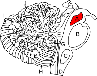
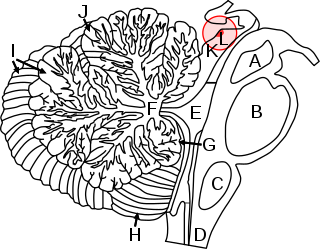
The brainstem is the stalk-like part of the brain that interconnects the cerebrum and diencephalon with the spinal cord. In the human brain, the brainstem is composed of the midbrain, the pons, and the medulla oblongata. The midbrain is continuous with the thalamus of the diencephalon through the tentorial notch.

The visual system is the physiological basis of visual perception. The system detects, transduces and interprets information concerning light within the visible range to construct an image and build a mental model of the surrounding environment. The visual system is associated with the eye and functionally divided into the optical system and the neural system.

In neuroanatomy, the lateral geniculate nucleus is a structure in the thalamus and a key component of the mammalian visual pathway. It is a small, ovoid, ventral projection of the thalamus where the thalamus connects with the optic nerve. There are two LGNs, one on the left and another on the right side of the thalamus. In humans, both LGNs have six layers of neurons alternating with optic fibers.

The midbrain or mesencephalon is the rostral-most portion of the brainstem connecting the diencephalon and cerebrum with the pons. It consists of the cerebral peduncles, tegmentum, and tectum.

In neuroanatomy, the medial lemniscus, also known as Reil's band or Reil's ribbon, is a large ascending bundle of heavily myelinated axons that decussate (cross) in the brainstem, specifically in the medulla oblongata. The medial lemniscus is formed by the crossings of the internal arcuate fibers. The internal arcuate fibers are composed of axons of nucleus gracilis and nucleus cuneatus. The axons of the nucleus gracilis and nucleus cuneatus in the medial lemniscus have cell bodies that lie contralaterally.

In neuroanatomy, the superior colliculus is a structure lying on the roof of the mammalian midbrain. In non-mammalian vertebrates, the homologous structure is known as the optic tectum or optic lobe. The adjective form tectal is commonly used for both structures.
Axon guidance is a subfield of neural development concerning the process by which neurons send out axons to reach their correct targets. Axons often follow very precise paths in the nervous system, and how they manage to find their way so accurately is an area of ongoing research.

The abducens nucleus is the originating nucleus from which the abducens nerve (VI) emerges—a cranial nerve nucleus. This nucleus is located beneath the fourth ventricle in the caudal portion of the pons near the midline, medial to the sulcus limitans.

Retinotopy is the mapping of visual input from the retina to neurons, particularly those neurons within the visual stream. For clarity, 'retinotopy' can be replaced with 'retinal mapping', and 'retinotopic' with 'retinally mapped'.

Jerome Ysroael Lettvin, often known as Jerry Lettvin, was an American cognitive scientist, and Professor of Electrical and Bioengineering and Communications Physiology at the Massachusetts Institute of Technology (MIT). He is best known as the lead author of the paper, "What the Frog's Eye Tells the Frog's Brain" (1959), one of the most cited papers in the Science Citation Index. He wrote it along with Humberto Maturana, Warren McCulloch and Walter Pitts, and in the paper they gave special thanks and mention to Oliver Selfridge at MIT. Lettvin carried out neurophysiological studies in the spinal cord, made the first demonstration of "feature detectors" in the visual system, and studied information processing in the terminal branches of single axons. Around 1969, he originated the term "grandmother cell" to illustrate the logical inconsistency of the concept.

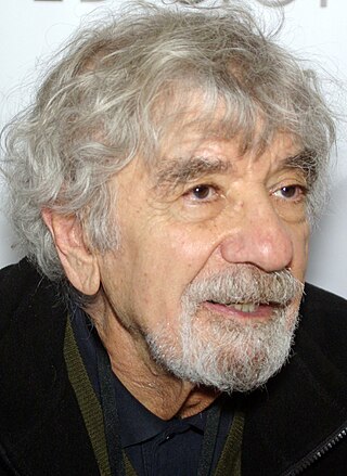
Humberto Maturana Romesín was a Chilean biologist and philosopher. Many consider him a member of a group of second-order cybernetics theoreticians such as Heinz von Foerster, Gordon Pask, Herbert Brün and Ernst von Glasersfeld, but in fact he was a biologist, scientist.
The Mauthner cells are a pair of big and easily identifiable neurons located in the rhombomere 4 of the hindbrain in fish and amphibians that are responsible for a very fast escape reflex. The cells are also notable for their unusual use of both chemical and electrical synapses.
Feature detection is a process by which the nervous system sorts or filters complex natural stimuli in order to extract behaviorally relevant cues that have a high probability of being associated with important objects or organisms in their environment, as opposed to irrelevant background or noise.

The growth cone is a highly dynamic structure of the developing neuron, changing directionality in response to different secreted and contact-dependent guidance cues; it navigates through the developing nervous system in search of its target. The migration of the growth cone is mediated through the interaction of numerous trophic and tropic factors; netrins, slits, ephrins and semaphorins are four well-studied tropic cues (Fig.1). The growth cone is capable of modifying its sensitivity to these guidance molecules as it migrates to its target; this sensitivity regulation is an important theme seen throughout development.

This article describes anatomical terminology that is used to describe the central and peripheral nervous systems - including the brain, brainstem, spinal cord, and nerves.

In anatomy a chiasm is the spot where two structures cross, forming an X-shape. Examples of chiasms are:
Target selection is the process by which axons selectively target other cells for synapse formation. Synapses are structures which enable electrical or chemical signals to pass between nerves. While the mechanisms governing target specificity remain incompletely understood, it has been shown in many organisms that a combination of genetic and activity-based mechanisms govern initial target selection and refinement. The process of target selection has multiple steps that include axon pathfinding when neurons extend processes to specific regions, cellular target selection when neurons choose appropriate partners in a target region from a multitude of potential partners, and subcellular target selection where axons often target particular regions of a partner neuron.

Raymond Michael ‘Mike’ Gaze FRS was a British neuroscientist.
References
- 1 2 "BIO254:Chemoaffinity". OpenWetWare. Retrieved 1 September 2011.
- 1 2 Ronald L. Meyer (1998). "Roger Sperry and his Chemoaffinity Hypothesis". Neuropsychologia. 36 (10): 957–980. doi:10.1016/S0028-3932(98)00052-9. PMID 9845045. S2CID 5665287.
- ↑ Ferry, Gorgina (10 June 1989). "The nervous system repairs to the network". New Scientist (1668). Retrieved 1 September 2011.(subscription required)
- ↑ Roger W. Sperry (1943). "Effect of 180 Degree Rotation of the Retinal Field on Visuomotor Coordination". The Journal of Experimental Zoology. 92 (3): 263–279. doi:10.1002/jez.1400920303.