Related Research Articles

Cholesteatoma is a destructive and expanding growth consisting of keratinizing squamous epithelium in the middle ear and/or mastoid process. Cholesteatomas are not cancerous as the name may suggest, but can cause significant problems because of their erosive and expansile properties. This can result in the destruction of the bones of the middle ear (ossicles), as well as growth through the base of the skull into the brain. They often become infected and can result in chronically draining ears. Treatment almost always consists of surgical removal.
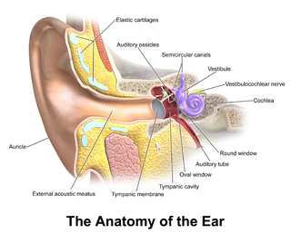
In the anatomy of humans and various other tetrapods, the eardrum, also called the tympanic membrane or myringa, is a thin, cone-shaped membrane that separates the external ear from the middle ear. Its function is to transmit sound from the air to the ossicles inside the middle ear, and then to the oval window in the fluid-filled cochlea. Hence, it ultimately converts and amplifies vibration in the air to vibration in cochlear fluid. The malleus bone bridges the gap between the eardrum and the other ossicles.

In anatomy, the temporomandibular joints (TMJ) are the two joints connecting the jawbone to the skull. It is a bilateral synovial articulation between the temporal bone of the skull above and the mandible below; it is from these bones that its name is derived. This joint is unique in that it is a bilateral joint that functions as one unit. Since the TMJ is connected to the mandible, the right and left joints must function together and therefore are not independent of each other.

The facial nerve, also known as the seventh cranial nerve, cranial nerve VII, or simply CN VII, is a cranial nerve that emerges from the pons of the brainstem, controls the muscles of facial expression, and functions in the conveyance of taste sensations from the anterior two-thirds of the tongue. The nerve typically travels from the pons through the facial canal in the temporal bone and exits the skull at the stylomastoid foramen. It arises from the brainstem from an area posterior to the cranial nerve VI and anterior to cranial nerve VIII.
Articles related to anatomy include:

The temporal bones are situated at the sides and base of the skull, and lateral to the temporal lobes of the cerebral cortex.

The otic ganglion is a small parasympathetic ganglion located immediately below the foramen ovale in the infratemporal fossa and on the medial surface of the mandibular nerve. It is functionally associated with the glossopharyngeal nerve and innervates the parotid gland for salivation.

The auriculotemporal nerve is a sensory branch of the mandibular nerve (CN V3) that runs with the superficial temporal artery and vein, and provides sensory innervation to parts of the external ear, scalp, and temporomandibular joint. The nerve also conveys post-ganglionic parasympathetic fibres from the otic ganglion to the parotid gland.

The internal auditory meatus is a canal within the petrous part of the temporal bone of the skull between the posterior cranial fossa and the inner ear.

In human anatomy, the mandibular canal is a canal within the mandible that contains the inferior alveolar nerve, inferior alveolar artery, and inferior alveolar vein. It runs obliquely downward and forward in the ramus, and then horizontally forward in the body, where it is placed under the alveoli and communicates with them by small openings.

The maxillary artery supplies deep structures of the face. It branches from the external carotid artery just deep to the neck of the mandible.

The squamous part of temporal bone, or temporal squama, forms the front and upper part of the temporal bone, and is scale-like, thin, and translucent.

The tympanic part of the temporal bone is a curved plate of bone lying below the squamous part of the temporal bone, in front of the mastoid process, and surrounding the external part of the ear canal.

The facial canal is a Z-shaped canal in the temporal bone of the skull. It extends between the internal acoustic meatus and stylomastoid foramen. It transmits the facial nerve.

The infratemporal fossa is an irregularly shaped cavity that is a part of the skull. It is situated below and medial to the zygomatic arch. It is not fully enclosed by bone in all directions. It contains superficial muscles, including the lower part of the temporalis muscle, the lateral pterygoid muscle, and the medial pterygoid muscle. It also contains important blood vessels such as the middle meningeal artery, the pterygoid plexus, and the retromandibular vein, and nerves such as the mandibular nerve (CN V3) and its branches.
The lesser petrosal nerve is the general visceral efferent (GVE) nerve conveying pre-ganglionic parasympathetic secretomotor fibers for the parotid gland from the tympanic plexus to the otic ganglion. It passes out of the tympanic cavity through the petrous part of the temporal bone into the middle cranial fossa of the cranial cavity, then exits the cranial cavity through its own canaliculus to reach the infratemporal fossa.

The mandibular fossa, also known as the glenoid fossa in some dental literature, is the depression in the temporal bone that articulates with the mandible.
The petrotympanic fissure is a fissure in the temporal bone that runs from the temporomandibular joint to the tympanic cavity.

The following outline is provided as an overview of and topical guide to human anatomy:
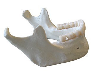
In anatomy, the mandible, lower jaw or jawbone is the bottom skeleton that makes up the lower half of the mouth in jawed vertebrates. In arthropods, the largest pair of appendages of their mouthparts are also named mandibles.
References
- 1 2 Lacout, Alexis; Marsot-Dupuch, Kathlyn; Smoker, Wendy R. K.; Lasjaunias, Pierre (2005-06-01). "Foramen Tympanicum, or Foramen of Huschke: Pathologic Cases and Anatomic CT Study". American Journal of Neuroradiology. 26 (6): 1317–1323. ISSN 0195-6108. PMID 15956489.
- ↑ Heffez, L.; Anderson, D.; Mafee, M. (1989). "Developmental defects of the tympanic plate: case reports and review of the literature". Journal of Oral and Maxillofacial Surgery. 47 (12): 1336–1340. doi:10.1016/0278-2391(89)90738-6. ISSN 0278-2391. PMID 2685214.
- ↑ Anand, V. T.; Latif, M. A.; Smith, W. P. (2000). "Defects of the external auditory canal: a new reconstruction technique". The Journal of Laryngology and Otology. 114 (4): 279–282. doi:10.1258/0022215001905553. ISSN 0022-2151. PMID 10845043.
- ↑ Tasar, Mustafa; Yetiser, Sertac (2003). "Congenital salivary fistula in the external auditory canal associated with chronic sialoadenitis and parotid cyst". Journal of Oral and Maxillofacial Surgery. 61 (9): 1101–1104. doi:10.1016/s0278-2391(03)00326-4. ISSN 0278-2391. PMID 12966489.
- ↑ Applebaum, E. L.; Berg, L. F.; Kumar, A.; Mafee, M. F. (1988). "Otologic complications following temporomandibular joint arthroscopy". The Annals of Otology, Rhinology, and Laryngology. 97 (6 Pt 1): 675–679. doi:10.1177/000348948809700618. ISSN 0003-4894. PMID 3202572.