Cranial nerves are the nerves that emerge directly from the brain, of which there are conventionally considered twelve pairs. Cranial nerves relay information between the brain and parts of the body, primarily to and from regions of the head and neck, including the special senses of vision, taste, smell, and hearing.

Bell's palsy is a type of facial paralysis that results in a temporary inability to control the facial muscles on the affected side of the face. In most cases, the weakness is temporary and significantly improves over weeks. Symptoms can vary from mild to severe. They may include muscle twitching, weakness, or total loss of the ability to move one or, in rare cases, both sides of the face. Other symptoms include drooping of the eyebrow, a change in taste, and pain around the ear. Typically symptoms come on over 48 hours. Bell's palsy can trigger an increased sensitivity to sound known as hyperacusis.

A smile is a facial expression formed primarily by flexing the muscles at the sides of the mouth. Some smiles include a contraction of the muscles at the corner of the eyes, an action known as a Duchenne smile. Among humans, a smile expresses delight, sociability, happiness, joy, or amusement. It is distinct from a similar but usually involuntary expression of anxiety known as a grimace. Although cross-cultural studies have shown that smiling is a means of communication throughout the world, there are large differences among different cultures, religions, and societies, with some using smiles to convey confusion, embarrassment or awkwardness.
A facial expression is one or more motions or positions of the muscles beneath the skin of the face. According to one set of controversial theories, these movements convey the emotional state of an individual to observers. Facial expressions are a form of nonverbal communication. They are a primary means of conveying social information between humans, but they also occur in most other mammals and some other animal species.

In neuroanatomy, the mandibular nerve (V3) is the largest of the three divisions of the trigeminal nerve, the fifth cranial nerve (CN V). Unlike the other divisions of the trigeminal nerve (ophthalmic nerve, maxillary nerve) which contain only afferent fibers, the mandibular nerve contains both afferent and efferent fibers. These nerve fibers innervate structures of the lower jaw and face, such as the tongue, lower lip, and chin. The mandibular nerve also innervates the muscles of mastication.

A facial is a family of skin care treatments for the face, including steam, exfoliation, extraction, creams, lotions, facial masks, peels, and massage. They are normally performed in beauty salons, but are also a common spa treatment. They are used for general skin health as well as for specific skin conditions. Types of facials include European facial, LED light therapy facials, hydrafacials and mini-facials.

The Facial Action Coding System (FACS) is a system to taxonomize human facial movements by their appearance on the face, based on a system originally developed by a Swedish anatomist named Carl-Herman Hjortsjö. It was later adopted by Paul Ekman and Wallace V. Friesen, and published in 1978. Ekman, Friesen, and Joseph C. Hager published a significant update to FACS in 2002. Movements of individual facial muscles are encoded by the FACS from slight different instant changes in facial appearance. It has proven useful to psychologists and to animators.

The lips are a horizontal pair of soft appendages attached to the jaws and are the most visible part of the mouth of many animals, including humans. Vertebrate lips are soft, movable and serve to facilitate the ingestion of food and the articulation of sound and speech. Human lips are also a somatosensory organ, and can be an erogenous zone when used in kissing and other acts of intimacy.

The corrugator supercilii muscle is a small, narrow, pyramidal muscle of the face. It arises from the medial end of the superciliary arch; it inserts into the deep surface of the skin of the eyebrow.

The nasalis muscle is a sphincter-like muscle of the nose. It has a transverse part and an alar part. It compresses the nasal cartilages, and can "flare" the nostrils. It can be used to test the facial nerve (VII), which supplies it.

The zygomaticus major muscle is a muscle of the face. It arises from either zygomatic arch (cheekbone); it inserts at the corner of the mouth. It is innervated by branches of the facial nerve.

The depressor anguli oris muscle is a facial muscle. It originates from the mandible and inserts into the angle of the mouth. It is associated with frowning, as it depresses the corner of the mouth.

The facial artery is a branch of the external carotid artery that supplies structures of the superficial face.

Facial toning, or facial exercise, is a type of cosmetic procedure or physical therapy tool which alters facial contours by means of increasing muscle tone and facial volume by promoting muscular hypertrophy, and preventing muscle loss due to aging or facial paralysis. Facial toning and exercise is therefore in part a technique to achieve facial rejuvenation by reducing wrinkles, sagging, and expression marks on the face and skin. As a physical therapy, facial toning is used for victims of stroke and forms of facial paralysis such as Bell’s palsy. Facial toning achieves this by performing facial muscle exercising. There are two types of facial toning exercises: active and passive face exercises.

The fascia lata is the deep fascia of the thigh. It encloses the thigh muscles and forms the outer limit of the fascial compartments of thigh, which are internally separated by the medial intermuscular septum and the lateral intermuscular septum. The fascia lata is thickened at its lateral side where it forms the iliotibial tract, a structure that runs to the tibia and serves as a site of muscle attachment.

The facial motor nucleus is a collection of neurons in the brainstem that belong to the facial nerve. These lower motor neurons innervate the muscles of facial expression and the stapedius.
The masseteric nerve is a nerve of the face. It is a branch of the mandibular nerve (CN V3). It passes through the mandibular notch to reach masseter muscle. It provides motor innervation the masseter muscle, and sensory innervation to the temporomandibular joint.

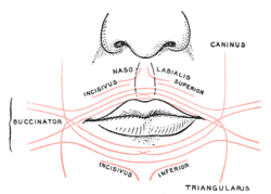
The facial muscles are a group of striated skeletal muscles supplied by the facial nerve that, among other things, control facial expression. These muscles are also called mimetic muscles. They are only found in mammals, although they derive from neural crest cells found in all vertebrates. They are the only muscles that attach to the dermis.
Central facial palsy is a symptom or finding characterized by paralysis or paresis of the lower half of one side of the face. It usually results from damage to upper motor neurons of the facial nerve.
The buccal fat pad is one of several encapsulated fat masses in the cheek. It is a deep fat pad located on either side of the face between the buccinator muscle and several more superficial muscles. The inferior portion of the buccal fat pad is contained within the buccal space. It should not be confused with the malar fat pad, which is directly below the skin of the cheek. It should also not be confused with jowl fat pads. It is implicated in the formation of hollow cheeks and the nasolabial fold, but not in the formation of jowls.