Type I collagen

Last updated
collagen, type I, alpha 1
Identifiers
SymbolCOL1A1
NCBI gene 1277
HGNC 2197
OMIM 120150
RefSeq NM_000088
UniProt P02452
Other data
Locus Chr. 17 q21.3-q22
Search for
Structures Swiss-model
Domains InterPro
collagen, type I, alpha 2
Identifiers
SymbolCOL1A2
Alt. symbolsOI4
NCBI gene 1278
HGNC 2198
OMIM 120160
RefSeq NM_000089
UniProt P08123
Other data
Locus Chr. 7 q21.3-22.1
Search for
Structures Swiss-model
Domains InterPro

Type I collagen is the most abundant collagen of the human body, consisting of around 90% of the body's total collagen in vertebrates. Due to this, it is also the most abundant protein type found in all vertebrates. Type I forms large, eosinophilic fibers known as collagen fibers, which make up most of the rope-like dense connective tissue in the body. [1] Collagen I itself is created by the combination of both a proalpha1 and a proalpha2 chain created by the COL1alpha1 and COL1alpha2 genes respectively. The Col I gene itself takes up a triple-helical conformation due to its Glycine-X-Y structure, x and y being any type of amino acid. Collagen can also be found in two different isoforms, either as a homotrimer or a heterotrimer, both of which can be found during different periods of development. Heterotrimers, in particular, play an important role in wound healing, [2] and are the dominant isoform found in the body. [2]

Contents

Type I collagen can be found in a myriad of different places in the body, mainly forming the matrix of connective tissues. It is present in scar tissue as well as tendons, ligaments, the endomysium of myofibrils, the organic part of bone, the dermis, the dentin, and organ capsules.

Formation

The creation process of type I Collagen begins with the production and the combination of two separate subunits, called the pro-alpha1(I) and pro-alpha2(I) chains. These pro-alpha chains are encoded by the COL1A1 and COL1A2 genes respectively and when combined produce type I pro-collagen. [3] This transcriptional process takes place within the cell's endoplasmic reticulum and must undergo post-translational modifications in order to make the final type I collagen product. [4] The procollagen complex is then modified by different enzyme proteinases which cleave N and C terminal pro-peptides that are present on either side of the molecule. This process occurs outside of the cellular membrane at which post processing, the molecules cross link and form a final type I collagen product.

Structure

Chemical Structure of Type I Collagen Collagen Type 1.png
Chemical Structure of Type I Collagen

Type I collagen has a triple-helical form which is caused by its amino acid composition. Its specific domain follows an order of G-X-Y In which the X and Y slots are occupied by any amino acid other than glycine however these slots are typically occupied by both hydroxyproline and proline, not in any particular order. [5] This specific conformation will end up being repeated and packed into a hexagonal structure in order to form collagen fibrils.

The molecular mass of type I collagen is 300,000 g/mol and assembles in one of two higher order molecular assemblies. It forms a large solid structure formed by strict and non-flexible protein interactions. [6] This large multi-protein structure is crucially held together by mainly hydrogen bonds and the fibrils conform to a typical diameter size between 25 - 400 nanometers in this fibril conformation. [5]    

Implications in Disease

Mutations in genes encoding collagen type 1 are known to cause a myriad of different conditions including:

Cardiac Valvular type Ehlers-Danlos Syndrome: This type of Ehlers-Danlos is caused by mutations within the COL1alpha2 gene, which is responsible for encoding the collagen pro-alpha2 chain. [7]

Vascular type Ehlers-Danlos Syndrome: Some patients with Vascular type Ehlers-Danlos, which is caused by mutations in COL3alpha1, are known to also have mutations in the COL1alpha1 gene. However the exact associations remain unknown. [8]

Athrochalasia type Ehlers-Danlos Syndrome: This type of Ehlers-Danlos is caused by the mutation of the COL1alpha1 and COL1alpha2 genes, which are responsible for encoding the proalpha1 and proalpha2 chains respectively.

Osteogenesis Imperfecta (types 1–4): Mutations in COL1alpha 1 and/or COL1alpha2 are known to cause several different types of Osteogenesis Imperfecta with the severity of said diseases being related to the type and frequency of the mutations occurring. [9] For further information on COL1's effect in this disease, see Collagen, type 1, alpha 1.

Caffey Disease: This condition is caused by a mutation in the COL1alpha gene that replaces arginine with cysteine at the 836 protein site. This particular mutation causes the fibrils of type I to vary greatly in size and shape. [10]

Clinical significance

See Collagen, type I, alpha 1#Clinical significance

Markers used to measure bone loss are not easily testable. Degradation of type I collagen releases metabolites that can be used to monitor resorption. [11]

See also

Related Research Articles

<span class="mw-page-title-main">Collagen</span> Most abundant structural protein in animals

Collagen is the main structural protein in the extracellular matrix found in the body's various connective tissues. As the main component of connective tissue, it is the most abundant protein in mammals, making up from 25% to 35% of the whole-body protein content. Collagen consists of amino acids bound together to form a triple helix of elongated fibril known as a collagen helix. It is mostly found in connective tissue such as cartilage, bones, tendons, ligaments, and skin. Vitamin C is vital for collagen synthesis, and Vitamin E improves the production of collagen.

<span class="mw-page-title-main">Osteogenesis imperfecta</span> Group of genetic disorders that mainly affect the bones

Osteogenesis imperfecta, colloquially known as brittle bone disease, is a group of genetic disorders that all result in bones that break easily. The range of symptoms—on the skeleton as well as on the body's other organs—may be mild to severe. Symptoms found in various types of OI include whites of the eye (sclerae) that are blue instead, short stature, loose joints, hearing loss, breathing problems and problems with the teeth. Potentially life-threatening complications, all of which become more common in more severe OI, include: tearing (dissection) of the major arteries, such as the aorta; pulmonary valve insufficiency secondary to distortion of the ribcage; and basilar invagination.

<span class="mw-page-title-main">Collagen, type II, alpha 1</span>

Collagen, type II, alpha 1 , also known as COL2A1, is a human gene that provides instructions for the production of the pro-alpha1(II) chain of type II collagen.

<span class="mw-page-title-main">ADAMTS2</span> Protein-coding gene in the species Homo sapiens

A disintegrin and metalloproteinase with thrombospondin motifs 2 (ADAM-TS2) also known as procollagen I N-proteinase is an enzyme that in humans is encoded by the ADAMTS2 gene.

<span class="mw-page-title-main">Collagen, type I, alpha 1</span> Mammalian protein found in humans

Collagen, type I, alpha 1, also known as alpha-1 type I collagen, is a protein that in humans is encoded by the COL1A1 gene. COL1A1 encodes the major component of type I collagen, the fibrillar collagen found in most connective tissues, including cartilage.

<span class="mw-page-title-main">Collagen, type XI, alpha 2</span> Protein found in humans

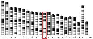
Collagen alpha-2(XI) chain is a protein that in humans is encoded by the COL11A2 gene.

<span class="mw-page-title-main">Dentinogenesis imperfecta</span> Medical condition

Dentinogenesis imperfecta (DI) is a genetic disorder of tooth development. It is inherited in an autosomal dominant pattern, as a result of mutations on chromosome 4q21, in the dentine sialophosphoprotein gene (DSPP). It is one of the most frequently occurring autosomal dominant features in humans. Dentinogenesis imperfecta affects an estimated 1 in 6,000-8,000 people.

Lysyl hydroxylases are alpha-ketoglutarate-dependent hydroxylases enzymes that catalyze the hydroxylation of lysine to hydroxylysine. Lysyl hydroxylases require iron and vitamin C as cofactors for their oxidation activity. It takes place following collagen synthesis in the cisternae (lumen) of the rough endoplasmic reticulum (ER). There are three lysyl hydroxylases (LH1-3) encoded in the human genome, namely: PLOD1, PLOD2 and PLOD3. From PLOD2 two splice variant can be expressed, where LH2b differs from LH2a by incorporating the small exon 13A. LH1 and LH3 hydroxylate lysyl residues in the collagen triple helix, whereas LH2b hydroxylates lysyl residues in the telopeptides of collagen. In addition to its hydroxylation activity, LH3 has glycosylation activity that produces either monosaccharide (Gal) or disaccharide (Glc-Gal) attached to collagen hydroxylysines.

<span class="mw-page-title-main">Sack–Barabas syndrome</span> Medical condition

Sack–Barabas syndrome is an older name for the medical condition vascular Ehlers–Danlos syndrome (vEDS). It affects the body's blood vessels and organs, making them prone to rupture.

<span class="mw-page-title-main">Collagen, type III, alpha 1</span>

Type III Collagen is a homotrimer, or a protein composed of three identical peptide chains (monomers), each called an alpha 1 chain of type III collagen. Formally, the monomers are called collagen type III, alpha-1 chain and in humans are encoded by the COL3A1 gene. Type III collagen is one of the fibrillar collagens whose proteins have a long, inflexible, triple-helical domain.

Type V collagen is a form of fibrillar collagen associated with classical Ehlers-Danlos syndrome. It is found within the dermal/epidermal junction, placental tissues, as well as in association with tissues containing type I collagen.

<span class="mw-page-title-main">Collagen, type V, alpha 1</span> Protein found in humans

Collagen alpha-1(V) chain is a protein that in humans is encoded by the COL5A1 gene.

<span class="mw-page-title-main">Collagen, type V, alpha 2</span> Protein found in humans

Collagen alpha-2(V) chain is a protein that in humans is encoded by the COL5A2 gene.

<span class="mw-page-title-main">Collagen, type I, alpha 2</span> Protein found in humans

Collagen alpha-2(I) chain is a protein that in humans is encoded by the COL1A2 gene.

<span class="mw-page-title-main">Collagen, type XI, alpha 1</span> Protein found in humans

Collagen alpha-1(XI) chain is a protein that in humans is encoded by the COL11A1 gene.

<span class="mw-page-title-main">Collagen, type VIII, alpha 1</span> Protein found in humans

Collagen alpha-1(VIII) chain is a protein that in humans is encoded by the COL8A1 gene.

<span class="mw-page-title-main">Collagen, type V, alpha 3</span> Protein found in humans

Collagen alpha-3(V) chain is a protein that in humans is encoded by the COL5A3 gene.

<span class="mw-page-title-main">CHST14</span> Protein-coding gene in the species Homo sapiens

Carbohydrate sulfotransferase 14 is an enzyme that in humans is encoded by the CHST14 gene.

<span class="mw-page-title-main">Bruck syndrome</span> Medical condition

Bruck syndrome is characterized as the combination of arthrogryposis multiplex congenita and osteogenesis imperfecta. Both diseases are uncommon, but concurrence is extremely rare which makes Bruck syndrome very difficult to research. Bruck syndrome is thought to be an atypical variant of osteogenesis imperfecta most resembling type III, if not its own disease. Multiple gene mutations associated with osteogenesis imperfecta are not seen in Bruck syndrome. Many affected individuals are within the same family, and pedigree data supports that the disease is acquired through autosomal recessive inheritance. Bruck syndrome has features of congenital contractures, bone fragility, recurring bone fractures, flexion joint and limb deformities, pterygia, short body height, and progressive kyphoscoliosis. Individuals encounter restricted mobility and pulmonary function. A reduction in bone mineral content and larger hydroxyapatite crystals are also detectable Joint contractures are primarily bilateral and symmetrical, and most prone to ankles. Bruck syndrome has no effect on intelligence, vision, or hearing.

<span class="mw-page-title-main">Daniel S. Greenspan</span> American biomedical scientist

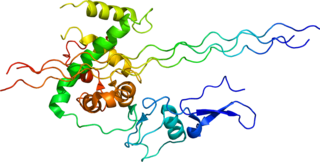
Daniel S. Greenspan is an American biomedical scientist, academic and researcher. He is Kellett professor of Cell and Regenerative Biology at the University of Wisconsin-Madison School of Medicine and Public Health. He has authored over 120 publications. His research has mainly focused on genes encoding proteins of the extracellular space and possible links between defects in such genes and human development and disease.

References

  1. "Collagen: What It Is, Types, Function & Benefits". Cleveland Clinic. Retrieved 2023-10-25.
  2. 1 2 Elena Makareeva (2014). "Collagen Structure, Folding and Function". Osteogenesis Imperfecta a Translational Approach to Brittle Bone Disease: 71–84. doi:10.1016/B978-0-12-397165-4.00007-1. ISBN   9780123971654.
  3. "COL1A2 gene". MedlinePlus Genetics. U.S. National Library of Medicine. Retrieved 2023-10-25.
  4. Canty-Laird EG, Lu Y, Kadler KE (January 2012). "Stepwise proteolytic activation of type I procollagen to collagen within the secretory pathway of tendon fibroblasts in situ". The Biochemical Journal. 441 (2): 707–717. doi:10.1042/BJ20111379. PMC   3430002 . PMID   21967573.
  5. 1 2 Naomi R, Ridzuan PM, Bahari H (August 2021). "Current Insights into Collagen Type I". Polymers. 13 (16): 2642. doi: 10.3390/polym13162642 . PMC   8399689 . PMID   34451183.
  6. Xia S, Chen Z, Shen C, Fu TM (September 2021). "Higher-order assemblies in immune signaling: supramolecular complexes and phase separation". Protein & Cell. 12 (9): 680–694. doi:10.1007/s13238-021-00839-6. PMC   8403095 . PMID   33835418.
  7. "Cardiac-Valvular Ehlers-Danlos Syndrome (cvEDS)". The Ehlers Danlos Society. Retrieved 2023-09-27.
  8. "Ehlers Danlos Syndromes - Symptoms, Causes, Treatment | NORD". rarediseases.org. Retrieved 2023-09-27.
  9. Gajko-Galicka A (2002). "Mutations in type I collagen genes resulting in osteogenesis imperfecta in humans". Acta Biochimica Polonica. 49 (2): 433–441. doi: 10.18388/abp.2002_3802 . PMID   12362985.
  10. "Caffey disease". MedlinePlus Genetics. U.S. National Library of Medicine. Retrieved 2023-09-27.
  11. Ting KR, Brady JJ, Hameed A, Le G, Meiller J, Verburgh E, et al. (April 2016). "Clinical utility of C-terminal telopeptide of type 1 collagen in multiple myeloma". British Journal of Haematology. 173 (1): 82–88. doi: 10.1111/bjh.13928 . PMID   26787413.