Patellar tendinitis

Last updated

Patellar tendinitis
Other namesquadriceps tendinopathy, patellar tendinopathy, jumper's knee, patellar tendinosis, patellar tendinitis
Jumpers knee.jpg
Location of the pain in patellar tendinitis
Specialty Orthopedics, sports medicine
Symptoms Pain at the front of the knee [1]
Complications Patellar tendon rupture [2]
Risk factors Jumping sports, being overweight [1]
Diagnostic method Based on symptoms and examination [2]
Differential diagnosis Chondromalacia patella, Osgood-Schlatter disease, patellofemoral syndrome, infrapatellar bursitis [1] [2]
TreatmentRest, physical therapy [2]
Prognosis Recovery can be slow [2]
Frequency14% of athletes [1]

Patellar tendinitis, also known as jumper's knee, is an overuse injury of the tendon that straightens the knee. [1] Symptoms include pain in the front of the knee. [1] Typically the pain and tenderness is at the lower part of the kneecap, though the upper part may also be affected. [2] Generally there is no pain when the person is at rest. [2] Complications may include patellar tendon rupture. [2]

Contents

Risk factors include being involved in athletics and being overweight. [1] It is particularly common in athletes who are involved in jumping sports such as basketball and volleyball. [1] [2] Other risk factors include sex, age, occupation, and physical activity level. [3] It is increasingly more likely to be developed with increasing age. [3] The underlying mechanism involves small tears in the tendon connecting the kneecap with the shinbone. [2] Diagnosis is generally based on symptoms and examination. [2] Other conditions that can appear similar include infrapatellar bursitis, chondromalacia patella and patellofemoral syndrome. [1] [2]

Treatment often involves resting the knee and physical therapy. [2] Evidence for treatments, including rest, however is poor. [4] [5] Recovery can take months and persist over years. [6] [7] [8] [2] It is relatively common with about 14% of athletes currently affected; however research reflects that more than half of athletes with this injury end their careers as a result. [9] [8] [1] Males are more commonly affected than females. [2] The term "jumper's knee" was coined in 1973. [2]

Signs and symptoms

People report anterior knee pain, often with an aching quality. The symptom onset is insidious. Rarely is a discrete injury described. Usually, the problem is below the kneecap but it may also be above. Jumper's knee can be classified into 1 of 4 stages, as follows: [2]

Stage 1: Pain only after activity, without functional impairment

Stage 2: Pain during and after activity, although the person is still able to perform satisfactorily in his or her sport

Stage 3: Prolonged pain during and after activity, with increasing difficulty in performing at a satisfactory level

Stage 4: Complete tendon tear requiring surgical repair

It begins as inflammation in the patellar tendon where it attaches to the patella and may progress by tearing or degenerating the tendon. People present with an ache over the patella tendon. Magnetic resonance imaging can reveal edema (increased T2 signal intensity) in the proximal aspect of the patellar tendon.[ citation needed ]

Causes

It is an overuse injury from repetitive overloading of the extensor mechanism of the knee. The microtears exceed the body's healing mechanism unless the activity is stopped.[ citation needed ]

Among the risk factors for patellar tendonitis are low ankle dorsiflexion, weak gluteal muscles, and muscle tightness, particularly in the calves, quadriceps muscle, and hamstrings. [10]

It may be associated with stiff ankle movement and ankle sprains. [11] [12]

Diagnosis

Diagnosis is generally based on symptoms and a physical examination. [2] Ultrasound or magnetic resonance imaging may help clarify how severe the problem is. [2]

Having a clinical diagnosis is the preferred way to diagnose patellar tendonitis, due to ultrasonographic abnormality. [3]

Treatment

Evidence for treatment is poor. [4] In the early stages rest, ice, compression, and elevation may be tried. Tentative evidence supports exercises involving eccentric muscle contractions of the quadriceps on a decline board. [13] Specific exercises and stretches to strengthen the muscles and tendons may be recommended, e.g. cycling or swimming. Use of a strap for jumper's knee and suspension inlays for shoes may also reduce the problems. Corticosteroid injections and NSAIDs are generally recommended. [2] [ dubious ]

Procedures

Dry needling, sclerosing injections, platelet-rich plasma, extracorporeal shock wave treatment (ESWT), and heat therapy have been tried. [2]

According to a study comparing ESWT to conservative treatment, it was found with low certainty that ESWT has a large treatment effect to reduce short term pain. [3]

Surgery

Surgery may be tried if other measures fail. [2] This may involve removal of myxoid degeneration in the tendon. This is reserved for people with severe pain for 6–12 months despite conservative measures. Novel treatment modalities targeting the abnormal blood vessel growth which occurs in the condition are currently being investigated.[ citation needed ] Knee operations in most cases have no better effects than exercise programs.[ citation needed ]

Epidemiology

It is relatively common with about 14% of athletes currently affected. [1] Males are more commonly affected than females. [2]

Related Research Articles

<span class="mw-page-title-main">Knee</span> Leg joint in primates

In humans and other primates, the knee joins the thigh with the leg and consists of two joints: one between the femur and tibia, and one between the femur and patella. It is the largest joint in the human body. The knee is a modified hinge joint, which permits flexion and extension as well as slight internal and external rotation. The knee is vulnerable to injury and to the development of osteoarthritis.

<span class="mw-page-title-main">Tendinopathy</span> Inflammation of the tendon

Tendinopathy is a type of tendon disorder that results in pain, swelling, and impaired function. The pain is typically worse with movement. It most commonly occurs around the shoulder, elbow, wrist, hip, knee, or ankle.

<span class="mw-page-title-main">Patella</span> Kneecap, bone covering knee joint

The patella, also known as the kneecap, is a flat, rounded triangular bone which articulates with the femur and covers and protects the anterior articular surface of the knee joint. The patella is found in many tetrapods, such as mice, cats, birds and dogs, but not in whales, or most reptiles.

<span class="mw-page-title-main">Achilles tendon</span> Tendon at the back of the lower leg

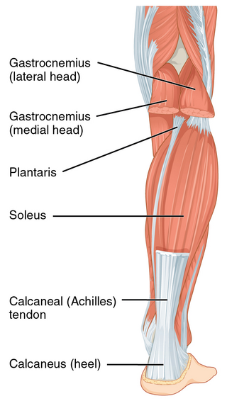
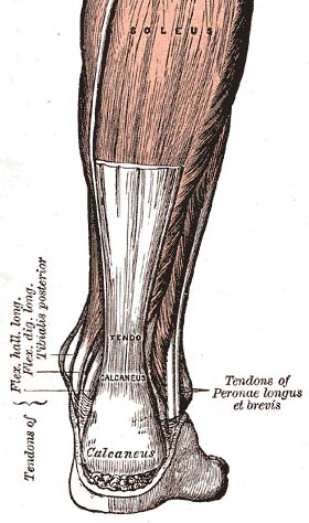
The Achilles tendon or heel cord, also known as the calcaneal tendon, is a tendon at the back of the lower leg, and is the thickest in the human body. It serves to attach the plantaris, gastrocnemius (calf) and soleus muscles to the calcaneus (heel) bone. These muscles, acting via the tendon, cause plantar flexion of the foot at the ankle joint, and flexion at the knee.

<span class="mw-page-title-main">Luxating patella</span> Medical condition

A luxating patella, sometimes called a trick knee, is a condition in which the patella, or kneecap, dislocates or moves out of its normal location. It can be associated with damage to the anterior cruciate ligament.

<span class="mw-page-title-main">Achilles tendinitis</span> Medical condition of the ankle and heel

Achilles tendinitis, also known as achilles tendinopathy, occurs when the Achilles tendon, found at the back of the ankle, becomes sore. Achilles tendinopathy is accompanied by alterations in the tendon's structure and mechanical properties. The most common symptoms are pain and swelling around the affected tendon. The pain is typically worse at the start of exercise and decreases thereafter. Stiffness of the ankle may also be present. Onset is generally gradual.

<span class="mw-page-title-main">Osgood–Schlatter disease</span> Inflammation of the patellar ligament

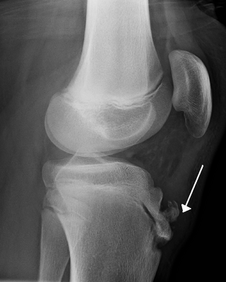
Osgood–Schlatter disease (OSD) is inflammation of the patellar ligament at the tibial tuberosity (apophysitis) usually affecting adolescents during growth spurts. It is characterized by a painful bump just below the knee that is worse with activity and better with rest. Episodes of pain typically last a few weeks to months. One or both knees may be affected and flares may recur.

<span class="mw-page-title-main">Chondromalacia patellae</span> Medical condition

Chondromalacia patellae is an inflammation of the underside of the patella and softening of the cartilage.

A negative repetition is the repetition of a technique in weight lifting in which the lifter performs the eccentric phase of a lift. Instead of pressing the weight up slowly, in proper form, a spotter generally aids in the concentric, or lifting, portion of the repetition while the lifter slowly performs the eccentric phase for 3–6 seconds.

<span class="mw-page-title-main">Patellar tendon rupture</span> Medical condition

Patellar tendon rupture is a tear of the tendon that connects the knee cap (patella) to the tibia. Often there is sudden onset of pain and walking is difficult. In a complete rupture, the ability to extend that knee is decreased. A pop may be felt when it occurs.

<span class="mw-page-title-main">Patella fracture</span> Medical condition

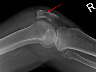
A patella fracture is a break of the kneecap. Symptoms include pain, swelling, and bruising to the front of the knee. A person may also be unable to walk. Complications may include injury to the tibia, femur, or knee ligaments.

<span class="mw-page-title-main">Unhappy triad</span> Medical condition of the knee

The unhappy triad, also known as a blown knee among other names, is an injury to the anterior cruciate ligament, medial collateral ligament, and meniscus. Analysis during the 1990s indicated that this 'classic' O'Donoghue triad is actually an unusual clinical entity among athletes with knee injuries. Some authors mistakenly believe that in this type of injury, "combined anterior cruciate and medial collateral ligament disruptions that were incurred during athletic endeavors" always present with concomitant medial meniscus injury. However, the 1990 analysis showed that lateral meniscus tears are more common than medial meniscus tears in conjunction with sprains of the ACL.

Arthrofibrosis has been described in most joints like knee, hip, ankle, foot joints, shoulder, elbow, wrist, hand joints as well as spinal vertebrae. It can occur after injury or surgery or may arise without an obvious cause. There is excessive scar tissue formation within the joint and/or surrounding soft tissues leading to painful restriction of joint motion that persists despite physical therapy and rehabilitation. The scar tissue may be located inside the knee joint or may involve the soft tissue structures around the knee joint, or both locations.

<span class="mw-page-title-main">Quadriceps tendon</span>

In human anatomy, the quadriceps tendon works with the quadriceps muscle to extend the leg. All four parts of the quadriceps muscle attach to the shin via the patella, where the quadriceps tendon becomes the patellar ligament. It attaches the quadriceps to the top of the patella, which in turn is connected to the shin from its bottom by the patellar ligament. A tendon connects muscle to bone, while a ligament connects bone to bone.

<span class="mw-page-title-main">Patellofemoral pain syndrome</span> Medical condition

Patellofemoral pain syndrome is knee pain as a result of problems between the kneecap and the femur. The pain is generally in the front of the knee and comes on gradually. Pain may worsen with sitting, excessive use, or climbing and descending stairs.

<span class="mw-page-title-main">Patellar dislocation</span> Medical condition

A patellar dislocation is a knee injury in which the patella (kneecap) slips out of its normal position. Often the knee is partly bent, painful and swollen. The patella is also often felt and seen out of place. Complications may include a patella fracture or arthritis.

<span class="mw-page-title-main">Knee pain</span> Medical condition

Knee pain is pain in or around the knee.

<span class="mw-page-title-main">Quadriceps tendon rupture</span> Tear of the tendon that runs from the quadriceps muscle to the top of the knee cap

A quadriceps tendon rupture is a tear of the tendon that runs from the quadriceps muscle to the top of the knee cap.

<span class="mw-page-title-main">Volleyball injuries</span>

Volleyball is a game played between two opposing sides, with six players on each team, where the players use mainly their hands to hit the ball over a net and try to make the ball land on the opposing team's side of the court. Volleyball is played by over 800 million people worldwide, making it one of the most popular sports in the world. Volleyball has some risks involved with it because there are some injuries which occur to players that are quite common; these include ankle injuries, shoulder injuries, foot injuries and knee injuries.

Muscle strain is one of the most common injuries in tennis. When an isolated large-energy appears during the muscle contraction and at the same time, bodyweight applies huge amounts of pressure to the lengthened muscle, which can result in the occurrence of muscle strain. Inflammation and bleeding are triggered when muscle strain occur which resulted in redness, pain and swelling. Overuse is also common in tennis players from all levels. Muscle, cartilage, nerves, bursae, ligaments and tendons may be damaged from overuse. The repetitive use of a particular muscle without time for repair and recover in the most common case among the injury.

References

  1. 1 2 3 4 5 6 7 8 9 10 11 King D, Yakubek G, Chughtai M, Khlopas A, Saluan P, Mont MA, Genin J (February 2019). "Quadriceps tendinopathy: a review-part 1: epidemiology and diagnosis". Annals of Translational Medicine. 7 (4): 71. doi: 10.21037/atm.2019.01.58 . PMC   6409230 . PMID   30963066.
  2. 1 2 3 4 5 6 7 8 9 10 11 12 13 14 15 16 17 18 19 20 21 22 23 Santana JA, Sherman A (January 2019). Jumpers Knee. Treasure Island, FL: StatPearls Publishing. PMID   30422564.
  3. 1 2 3 4 Charles R, Fang L, Zhu R, Wang J (2023). "The effectiveness of shockwave therapy on patellar tendinopathy, Achilles tendinopathy, and plantar fasciitis: a systematic review and meta-analysis". Frontiers in Immunology. 14: 1193835. doi: 10.3389/fimmu.2023.1193835 . ISSN   1664-3224. PMC   10468604 . PMID   37662911.
  4. 1 2 Mendonça LM, Leite HR, Zwerver J, Henschke N, Branco G, Oliveira VC (6 June 2019). "How strong is the evidence that conservative treatment reduces pain and improves function in individuals with patellar tendinopathy? A systematic review of randomised controlled trials including GRADE recommendations". British Journal of Sports Medicine. 54 (2): bjsports–2018–099747. doi:10.1136/bjsports-2018-099747. PMID   31171514. S2CID   174810797.
  5. Saithna A, Gogna R, Baraza N, Modi C, Spencer S (30 November 2012). "Eccentric Exercise Protocols for Patella Tendinopathy: Should we Really be Withdrawing Athletes from Sport? A Systematic Review". The Open Orthopaedics Journal. 6 (1): 553–557. doi: 10.2174/1874325001206010553 . ISSN   1874-3250. PMC   3522085 . PMID   23248727.
  6. Nuhmani S, Muaidi QI: Patellar tendinopathy: a review of literature. J Clin Diagn Res 2018;12. doi : 10.7860/JCDR/2018/35797.11605.
  7. Cummings K, Skinner L, Cushman DM: "Patellar tendinopathy in athletes". Curr Phys Med Rehabil Rep 2019;7:227–36
  8. 1 2 Plinsinga M.L., Meeus M., Brink M., Heugen N., Van Wilgen P. "Evidence of Widespread Mechanical Hyperalgesia but Not Exercise-Induced Analgesia in Athletes with Mild Patellar Tendinopathy Compared with Pain-Free Matched Controls: A Blinded Exploratory Study". Am. J. Phys. Med. Rehabil. 2021;100(10):946-951. doi:10.1097/PHM.0000000000001673
  9. Kettunen JA, Kvist M, Alanen E, et al: Long-term prognosis for Jumper's knee in male athletes: a prospective follow-up study. Am J Sports Med 2002;30:689–92
  10. Koban M (2013). Beating Patellar Tendonitis. CreateSpace Independent Publishing Platform. pp. 20–25. ISBN   978-1-4910-4973-0.
  11. Marcus A (7 October 2011). "Stiff ankles tied to young athletes' painful knees". Reuters.
  12. Backman LJ, Danielson P (December 2011). "Low range of ankle dorsiflexion predisposes for patellar tendinopathy in junior elite basketball players: a 1-year prospective study". The American Journal of Sports Medicine. 39 (12): 2626–33. doi:10.1177/0363546511420552. PMID   21917610. S2CID   39755525.
  13. Visnes H, Bahr R (April 2007). "The evolution of eccentric training as treatment for patellar tendinopathy (jumper's knee): a critical review of exercise programmes". British Journal of Sports Medicine. 41 (4): 217–23. doi:10.1136/bjsm.2006.032417. PMC   2658948 . PMID   17261559.