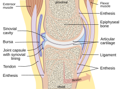
It is a type of enthesopathy, meaning any pathologic condition of the entheses, with or without inflammation. There are some cases of isolated, primary enthesitis which are very poorly studied and understood. It is known to be associated with other autoimmune diseases, like spondyloarthropathies and psoriasis (thought to often precede psoriatic arthritis). A common autoimmune enthesitis is at the heel, where the Achilles tendon attaches to the calcaneus.
Early clinical manifestations are an aching sensation akin to "working out too much", and it gets better with activity. It is worse in the morning (after sleeping and not moving). The muscle insertion hurts very focally as it joins into the bone, but there is little to no pain at all with passive motion.
Symptoms include multiple points of tenderness at the heel, tibial tuberosity, iliac crest, and other tendon insertion sites.
Diagnosis
Sagittal magnetic resonance images of ankle region: psoriatic arthritis. (a) Short tau inversion recovery (STIR) image, showing high signal intensity at the Achilles tendon insertion (enthesitis, thick arrow) and in the synovium of the ankle joint (synovitis, long thin arrow). Bone marrow oedema is seen at the tendon insertion (short thin arrow). (b, c) T1 weighted images of a different section of the same patient, before (panel b) and after (panel c) intravenous contrast injection, confirm inflammation (large arrow) at the enthesis and reveal bone erosion at tendon insertion (short thin arrows).
↑ Hendrix CL (2005). "Calcaneal apophysitis (Sever disease)". Clinics in Podiatric Medicine and Surgery. 22 (1): 55–62, vi. doi:10.1016/j.cpm.2004.08.011. PMID15555843.
↑ "Tendinitis". National Institute of Arthritis and Musculoskeletal and Skin Diseases. 12 April 2017. Archived from the original on October 4, 2017. Retrieved 18 November 2018.
This page is based on this Wikipedia article Text is available under the CC BY-SA 4.0 license; additional terms may apply. Images, videos and audio are available under their respective licenses.