Interphalangeal joints of the hand

Last updated
Joints of the hand
DIP, PIP and MCP joints of hand.jpg
The DIP, PIP and MCP joints of the hand:
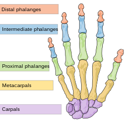
Scheme human hand bones-en.svg
Human hand bones
Details
Identifiers
Latin articulationes interphalangeae manus, articulationes digitorum manus
TA98 A03.5.11.601
TA2 1839
FMA 35285
Anatomical terminology

The interphalangeal joints of the hand are the hinge joints between the phalanges of the fingers that provide flexion towards the palm of the hand.

Contents

There are two sets in each finger (except in the thumb, which has only one joint):

Anatomically, the proximal and distal interphalangeal joints are very similar. There are some minor differences in how the palmar plates are attached proximally and in the segmentation of the flexor tendon sheath, but the major differences are the smaller dimension and reduced mobility of the distal joint. [1]

Joint structure

Joints of the hand, X-ray 814 Radiograph of Hand.jpg
Joints of the hand, X-ray
Interphalangeal ligaments and phalanges. Right hand. Deep dissection. Posterior (dorsal) view. Slide1tyty.JPG
Interphalangeal ligaments and phalanges. Right hand. Deep dissection. Posterior (dorsal) view.

The PIP joint exhibits great lateral stability. Its transverse diameter is greater than its antero-posterior diameter and its thick collateral ligaments are tight in all positions during flexion, contrary to those in the metacarpophalangeal joint. [1]

Dorsal structures

The capsule, extensor tendon, and skin are very thin and lax dorsally, allowing for both phalanx bones to flex more than 100° until the base of the middle phalanx makes contact with the condylar notch of the proximal phalanx. [1]

At the level of the PIP joint the extensor mechanism splits into three bands. The central slip attaches to the dorsal tubercle of the middle phalanx near the PIP joint. The pair of lateral bands, to which contribute the extensor tendons, continue past the PIP joint dorsally to the joint axis. These three bands are united by a transverse retinacular ligament, which runs from the palmar border of the lateral band to the flexor sheath at the level of the joint and which prevents dorsal displacement of that lateral band. On the palmar side of the joint axis of motion, lies the oblique retinacular ligament [of Landsmeer] which stretches from the flexor sheath over the proximal phalanx to the terminal extensor tendon. In extension, the oblique ligament prevents passive DIP flexion and PIP hyperextension as it tightens and pulls the terminal extensor tendon proximally. [2]

Palmar structures

In contrast, on the palmar side, a thick ligament prevents hyperextension. The distal part of the palmar ligament, called the palmar plate, is 2 to 3 millimetres (0.079 to 0.118 in) thick and has a fibrocartilaginous structure. The presence of chondroitin and keratan sulfate in the dorsal and palmar plates is important in resisting compression forces against the condyles of the proximal phalanx. Together these structures protect the tendons passing in front and behind the joint. These tendons can sustain traction forces thanks to their collagen fibers. [1]

Palmar ligament

Metacarpophalangeal joint and joints of digit. Palmar aspect. Palmar ligament labelled as volar ligament. Gray337.png
Metacarpophalangeal joint and joints of digit. Palmar aspect. Palmar ligament labelled as volar ligament.

The palmar ligament is thinner and more flexible in its central-proximal part. On both sides it is reinforced by the so-called check rein ligaments. The accessory collateral ligaments (ACL) originate at the proximal phalanx and are inserted distally at the base of the middle phalanx below the collateral ligaments. [1]

The accessory ligament and the proximal margin of the palmar plate are flexible and fold back upon themselves during flexion. The flexor tendon sheaths are firmly attached to the proximal and middle phalanges by annular pulleys A2 and A4, while the A3 pulley and the proximal fibres of the C1 ligament attach the sheaths to the mobile volar ligament at the PIP joint. During flexion this arrangement produces a space at the neck of the proximal phalanx which is filled by the folding palmar plate. [2]

The palmar plate is supported by a ligament on either side of the joint called the collateral ligaments, which prevent deviation of the joint from side to side. The ligaments can partially or fully tear and can avulse with a small fracture fragment when the finger is forced backwards into hyperextension. This is called a "palmar plate, or volar plate injury". [3]

The palmar plate forms a semi-rigid floor and the collateral ligaments the walls in a mobile box which moves together with the distal part of the joint and provides stability to the joint during its entire range of motion. Because the palmar plate adheres to the flexor digitorum superficialis near the distal attachment of the muscle, it also increases the moment of flexor action. In the PIP joint, extension is more limited because of the two so called check-rein ligaments, which attach the palmar plate to the proximal phalanx. [2]

Movements

The only movements permitted in the interphalangeal joints are flexion and extension.

The muscles generating these movements are:

Location Flexion Extension
fingers the flexor digitorum profundus acting on the proximal and distal joints, and the flexor digitorum superficialis acting on the proximal joints mainly by the lumbricals and interossei, the long extensors having little or no action upon these joints
thumb the flexor pollicis longus the extensor pollicis longus

The relative length of the digit varies during motion of the IP joints. The length of the palmar aspect decreases during flexion while the dorsal aspect increases by about 24 mm. The useful range of motion of the PIP joint is 30–70°, increasing from the index finger to the little finger. During maximum flexion the base of the middle phalanx is firmly pressed into the retrocondylar recess of the proximal phalanx, which provides maximum stability to the joint. The stability of the PIP joint is dependent of the tendons passing around it. [2]

Clinical significance

Rheumatoid arthritis generally spares the distal interphalangeal joints. [4] Therefore, arthritis of the distal interphalangeal joints strongly suggests the presence of osteoarthritis or psoriatic arthritis. [5]

See also

Related Research Articles

<span class="mw-page-title-main">Carpal bones</span> Eight small bones that make up the wrist (or carpus) that connects the hand to the forearm

The carpal bones are the eight small bones that make up the wrist that connects the hand to the forearm. The term "carpus" is derived from the Latin carpus and the Greek καρπός (karpós), meaning "wrist". In human anatomy, the main role of the wrist is to facilitate effective positioning of the hand and powerful use of the extensors and flexors of the forearm, and the mobility of individual carpal bones increase the freedom of movements at the wrist.

<span class="mw-page-title-main">Metacarpal bones</span> Bones of hand

In human anatomy, the metacarpal bones or metacarpus form the intermediate part of the skeletal hand located between the phalanges of the fingers and the carpal bones of the wrist, which forms the connection to the forearm. The metacarpal bones are analogous to the metatarsal bones in the foot.

<span class="mw-page-title-main">Upper limb</span> Consists of the arm, forearm, and hand

The upper limbs or upper extremities are the forelimbs of an upright-postured tetrapod vertebrate, extending from the scapulae and clavicles down to and including the digits, including all the musculatures and ligaments involved with the shoulder, elbow, wrist and knuckle joints. In humans, each upper limb is divided into the arm, forearm and hand, and is primarily used for climbing, lifting and manipulating objects.

<span class="mw-page-title-main">Phalanx bone</span> Digital bone in the hands and feet of most vertebrates

The phalanges are digital bones in the hands and feet of most vertebrates. In primates, the thumbs and big toes have two phalanges while the other digits have three phalanges. The phalanges are classed as long bones.

<span class="mw-page-title-main">Extensor digitorum muscle</span>

The extensor digitorum muscle is a muscle of the posterior forearm present in humans and other animals. It extends the medial four digits of the hand. Extensor digitorum is innervated by the posterior interosseous nerve, which is a branch of the radial nerve.

In human anatomy, the extensor pollicis longus muscle (EPL) is a skeletal muscle located dorsally on the forearm. It is much larger than the extensor pollicis brevis, the origin of which it partly covers and acts to stretch the thumb together with this muscle.

<span class="mw-page-title-main">Palmar interossei muscles</span> Muscles between the metacarpals

In human anatomy, the palmar or volar interossei are three small, unipennate muscles in the hand that lie between the metacarpal bones and are attached to the index, ring, and little fingers. They are smaller than the dorsal interossei of the hand.

<span class="mw-page-title-main">Metacarpophalangeal joint</span>

The metacarpophalangeal joints (MCP) are situated between the metacarpal bones and the proximal phalanges of the fingers. These joints are of the condyloid kind, formed by the reception of the rounded heads of the metacarpal bones into shallow cavities on the proximal ends of the proximal phalanges. Being condyloid, they allow the movements of flexion, extension, abduction, adduction and circumduction at the joint.

<span class="mw-page-title-main">Dorsal interossei of the hand</span> Muscles between the metacarpals

In human anatomy, the dorsal interossei (DI) are four muscles in the back of the hand that act to abduct (spread) the index, middle, and ring fingers away from hand's midline and assist in flexion at the metacarpophalangeal joints and extension at the interphalangeal joints of the index, middle and ring fingers.

<span class="mw-page-title-main">Boutonniere deformity</span> Medical condition

Boutonniere deformity is a deformed position of the fingers or toes, in which the joint nearest the knuckle is permanently bent toward the palm while the farthest joint is bent back away. Causes include injury, inflammatory conditions like rheumatoid arthritis, and genetic conditions like Ehlers-Danlos syndrome.

<span class="mw-page-title-main">Interphalangeal joints of the foot</span>

The interphalangeal joints of the foot are between the phalanx bones of the toes in the feet.

<span class="mw-page-title-main">Extensor expansion</span> Special connective attachments by which the extensor tendons insert into the phalanges

An extensor expansion is the special connective attachments by which the extensor tendons insert into the phalanges.

<span class="mw-page-title-main">Annular ligaments of fingers</span>

In human anatomy, the annular ligaments of the fingers, often referred to as A pulleys, are the annular part of the fibrous sheathes of the fingers. Four or five such annular pulleys, together with three cruciate pulleys, form a fibro-osseous tunnel on the palmar aspect of the hand through which passes the deep and superficial flexor tendons. The annular and cruciate ligaments serve to govern the flexor mechanism of the hand and wrist, providing critical constraints to the flexor tendons to prevent bowstringing upon contraction and excursion of extrinsic flexor musculo-tendinous units.

<span class="mw-page-title-main">Jammed finger</span> Medical condition

Jammed finger is a colloquialism referring to a variety of injuries to the joints of the fingers, resulting from axial loading beyond that which the ligaments can withstand. Common parts of the finger susceptible to this type of injury are ligaments, joints, and bones. The severity of the damage to the finger increases with the magnitude of the force exerted by the external object on the fingertip. Toes may become jammed as well, with similar results.

<span class="mw-page-title-main">Collateral ligaments of metacarpophalangeal joints</span>

In human anatomy, the radial (RCL) and ulnar (UCL) collateral ligaments of the metacarpophalangeal joints (MCP) of the hand are the primary stabilisers of the MCP joints. They have two parts: the cord-like collateral ligaments proper located more dorsally and the accessory collateral ligaments located more volarly. They enable us to spread our fingers with an open hand but not with the hand closed into a fist.

<span class="mw-page-title-main">Palmar plate</span>

In the human hand, palmar or volar plates are found in the metacarpophalangeal (MCP) and interphalangeal (IP) joints, where they reinforce the joint capsules, enhance joint stability, and limit hyperextension. The plates of the MCP and IP joints are structurally and functionally similar, except that in the MCP joints they are interconnected by a deep transverse ligament. In the MCP joints, they also indirectly provide stability to the longitudinal palmar arches of the hand. The volar plate of the thumb MCP joint has a transverse longitudinal rectangular shape, shorter than those in the fingers.

In the human foot, the plantar or volar plates are fibrocartilaginous structures found in the metatarsophalangeal (MTP) and interphalangeal (IP) joints. The anatomy and composition of the plantar plates are similar to the palmar plates in the metacarpophalangeal (MCP) and interphalangeal joints in the hand; the proximal origin is thin but the distal insertion is stout. Due to the weight-bearing nature of the human foot, the plantar plates are exposed to extension forces not present in the human hand.

<span class="mw-page-title-main">Swan neck deformity</span> Medical condition

Swan neck deformity is a deformed position of the finger, in which the joint closest to the fingertip is permanently bent toward the palm while the nearest joint to the palm is bent away from it. It is commonly caused by injury, hypermobility or inflammatory conditions like rheumatoid arthritis or sometimes familial.

<span class="mw-page-title-main">Hand</span> Extremity at the end of an arm or forelimb

A hand is a prehensile, multi-fingered appendage located at the end of the forearm or forelimb of primates such as humans, chimpanzees, monkeys, and lemurs. A few other vertebrates such as the koala are often described as having "hands" instead of paws on their front limbs. The raccoon is usually described as having "hands" though opposable thumbs are lacking.

<span class="mw-page-title-main">Extrinsic extensor muscles of the hand</span>

The extrinsic extensor muscles of the hand are located in the back of the forearm and have long tendons connecting them to bones in the hand, where they exert their action. Extrinsic denotes their location outside the hand. Extensor denotes their action which is to extend, or open flat, joints in the hand. They include the extensor carpi radialis longus (ECRL), extensor carpi radialis brevis (ECRB), extensor digitorum (ED), extensor digiti minimi (EDM), extensor carpi ulnaris (ECU), abductor pollicis longus (APL), extensor pollicis brevis (EPB), extensor pollicis longus (EPL), and extensor indicis (EI).

References

  1. 1 2 3 4 5 Lluch 1997 , pp. 259–60
  2. 1 2 3 4 Brüser & Gilbert 1999 , pp. 158–60
  3. "Hand Therapy - Volar Plate Injury".
  4. Victoria Ruffing, Clifton O. Bingham. "Rheumatoid Arthritis Signs and Symptoms". Johns Hopkins Hospital . Retrieved 2017-08-10. Updated: April 4, 2017
  5. PJW Venables, Ravinder N Maini. "Diagnosis and differential diagnosis of rheumatoid arthritis". UpToDate . Retrieved 2017-08-10. Last updated: Jan 22, 2016.

Further reading

PD-icon.svgThis article incorporates text in the public domain from page 333 of the 20th edition of Gray's Anatomy (1918)