Spinal nerve root may refer to:
| This article includes a list of related items that share the same name (or similar names). If an internal link incorrectly led you here, you may wish to change the link to point directly to the intended article. |
Spinal nerve root may refer to:
| This article includes a list of related items that share the same name (or similar names). If an internal link incorrectly led you here, you may wish to change the link to point directly to the intended article. |

The peripheral nervous system (PNS) is one of two components that make up the nervous system of bilateral animals, with the other part being the central nervous system (CNS). The PNS consists of the nerves and ganglia outside the brain and spinal cord. The main function of the PNS is to connect the CNS to the limbs and organs, essentially serving as a relay between the brain and spinal cord and the rest of the body. Unlike the CNS, the PNS is not protected by the vertebral column and skull, or by the blood–brain barrier, which leaves it exposed to toxins and mechanical injuries.

A spinal nerve is a mixed nerve, which carries motor, sensory, and autonomic signals between the spinal cord and the body. In the human body there are 31 pairs of spinal nerves, one on each side of the vertebral column. These are grouped into the corresponding cervical, thoracic, lumbar, sacral and coccygeal regions of the spine. There are eight pairs of cervical nerves, twelve pairs of thoracic nerves, five pairs of lumbar nerves, five pairs of sacral nerves, and one pair of coccygeal nerves. The spinal nerves are part of the peripheral nervous system.
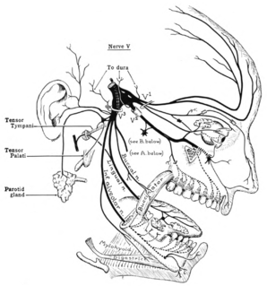
The trigeminal nerve (the fifth cranial nerve, or simply CN V) is a nerve responsible for sensation in the face and motor functions such as biting and chewing; it is the most complex of the cranial nerves. Its name ("trigeminal" = tri-, or three, and - geminus, or twin: thrice-twinned) derives from the fact that each of the two nerves (one on each side of the pons) has three major branches: the ophthalmic nerve (V1), the maxillary nerve (V2), and the mandibular nerve (V3). The ophthalmic and maxillary nerves are purely sensory, whereas the mandibular nerve supplies motor as well as sensory (or "cutaneous") functions. Adding to the complexity of this nerve is the fact that autonomic nerve fibers as well as special sensory fibers (taste) are contained within it.

In tetrapod anatomy, lumbar is an adjective that means of or pertaining to the abdominal segment of the torso, between the diaphragm and the sacrum.

A dorsal root ganglion is a cluster of neurons in a dorsal root of a spinal nerve. The cell bodies of sensory neurons known as first-order neurons are located in the dorsal root ganglia.

A dermatome is an area of skin that is mainly supplied by afferent nerve fibres from the dorsal root of any given spinal nerve. There are 8 cervical nerves , 12 thoracic nerves, 5 lumbar nerves and 5 sacral nerves. Each of these nerves relays sensation from a particular region of skin to the brain.
Radicular pain, or radiculitis, is pain "radiated" along the dermatome of a nerve due to inflammation or other irritation of the nerve root (radiculopathy) at its connection to the spinal column. A common form of radiculitis is sciatica – radicular pain that radiates along the sciatic nerve from the lower spine to the lower back, gluteal muscles, back of the upper thigh, calf, and foot as often secondary to nerve root irritation from a spinal disc herniation or from osteophytes in the lumbar region of the spine. Radiculitis indicates inflammation of the spinal nerve root, which may lead to pain in that nerve's distribution without weakness as opposed to radiculopathy. When the radiating pain is associated with numbness or weakness, the diagnosis is radiculopathy if the lesion is at the nerve root and myelopathy if at the spinal cord itself.

A pseudounipolar neuron is a type of neuron which has one extension from its cell body. This type of neuron contains an axon that has split into two branches; one branch travels to the peripheral nervous system and the other to the central nervous system. A single process arises from the cell body and then divides into an axon and a dendrite. They develop embryologically as bipolar in shape and are thus termed pseudounipolar instead of unipolar.
A myotome is the group of muscles that a single spinal nerve innervates. Similarly a dermatome is an area of skin that a single nerve innervates. In vertebrate embryonic development, a myotome is the part of a somite that develops into the muscles.
The dorsal root of spinal nerve is one of two "roots" which emerge from the spinal cord. It emerges directly from the spinal cord, and travels to the dorsal root ganglion. Nerve fibres with the ventral root then combine to form a spinal nerve. The dorsal root transmits sensory information, forming the afferent sensory root of a spinal nerve.
In anatomy and neurology, the ventral root or anterior root is the efferent motor root of a spinal nerve.
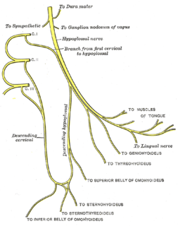
The ansa cervicalis is a loop of nerves that are part of the cervical plexus. It lies superficial to the internal jugular vein in the carotid triangle. Its name means "handle of the neck" in Latin.
A nerve root is the initial segment of a nerve leaving the central nervous system. Types include:
The dorsal ramus of spinal nerve is the posterior division of a spinal nerve. The dorsal ramus is the dorsal branch of a spinal nerve that forms from the dorsal root of the nerve after it emerges from the spinal cord. The spinal nerve is formed from the dorsal and ventral rami. The dorsal ramus carries information that supplies muscles and sensation to the human back.

The general visceral afferent (GVA) fibers conduct sensory impulses from the internal organs, glands, and blood vessels to the central nervous system. They are considered to be part of the visceral nervous system, which is closely related to the autonomic nervous system, but 'visceral nervous system' and 'autonomic nervous system' are not direct synonyms and care should be taken when using these terms. Unlike the efferent fibers of the autonomic nervous system, the afferent fibers are not classified as either sympathetic or parasympathetic.

Radiculopathy, also commonly referred to as pinched nerve, refers to a set of conditions in which one or more nerves are affected and do not work properly. This can result in pain, weakness, numbness, or difficulty controlling specific muscles.

The cranial root of accessory nerve is the smaller of the two portions of the accessory nerve. It is generally considered as a part of the vagus nerve and not part of the accessory nerve proper because the cranial component rapidly joins the vagus nerve and serves the same function as other vagal nerve fibers. Recently, the concept of a cranial root of the accessory nerve has been challenged by new neuroanatomical studies which found that an unambiguous cranial root was not present in the majority of the cases. However, a small study in 2007 followed by a substantially larger study published in 2012 both confirmed that the cranial root of the accessory nerve is commonly found in humans, matching traditional descriptions.
The spinal cord is a long, thin, tubular structure made up of nervous tissue, which extends from the medulla oblongata in the brainstem to the lumbar region of the vertebral column. It encloses the central canal of the spinal cord, which contains cerebrospinal fluid. The brain and spinal cord together make up the central nervous system (CNS). In humans, the spinal cord begins at the occipital bone, passing through the foramen magnum and entering the spinal canal at the beginning of the cervical vertebrae. The spinal cord extends down to between the first and second lumbar vertebrae, where it ends. The enclosing bony vertebral column protects the relatively shorter spinal cord. It is around 45 cm (18 in) in men and around 43 cm (17 in) long in women. The diameter of the spinal cord ranges from 13 mm in the cervical and lumbar regions to 6.4 mm in the thoracic area.

Spinal decompression is a surgical procedure intended to relieve pressure on the spinal cord or on one or more compressed nerve roots passing through or exiting the spinal column. Decompression of the spinal neural elements is a key component in treating spinal radiculopathy, myelopathy and claudication.

The ciliary ganglion is a parasympathetic ganglion located just behind the eye in the posterior orbit. Three types of axons enter the ciliary ganglion but only the preganglionic parasympathetic axons synapse there. The entering axons are arranged into three roots of the ciliary ganglion, which join enter the posterior surface of the ganglion.