Table of cranial nerves

Last updated

No.NameSensory, motor, or bothOrigin/TargetExited mannerFunction [1]
0 Terminal ? Lamina terminalis Located in the cribriform plate of the ethmoid bone.Animal research indicates that the terminal nerve is involved in the detection of pheromones. [2]
I Olfactory Purely sensory Telencephalon Located in the olfactory foramina in the cribriform plate of the ethmoid bone.Transmits the sense of smell from the nasal cavity. [3]
II Optic Sensory Retinal ganglion cells Located in the optic canal.Transmits visual signals from the retina of the eye to the brain. [4]
III Oculomotor Mainly motorAnterior aspect of Midbrain Located in the superior orbital fissure.Innervates the levator palpebrae superioris, superior rectus, medial rectus, inferior rectus, and inferior oblique, which collectively perform most eye movements. Also innervates the sphincter pupillae and the muscles of the ciliary body.
IV Trochlear MotorDorsal aspect of Midbrain Located in the superior orbital fissure.Innervates the superior oblique muscle, which depresses, abducts, and intorts the eyeball.
V Trigeminal Both sensory and motor Pons Three Parts:

V1 (ophthalmic nerve) is located in the superior orbital fissure V2 (maxillary nerve) is located in the foramen rotundum

V3 (mandibular nerve) is located in the foramen ovale.

Receives sensation from the face, mouth and nasal cavity, and innervates the muscles of mastication.
VI Abducens Mainly motorNuclei lying under the floor of the fourth ventricle

Pons

Located in the superior orbital fissure.Innervates the lateral rectus, which abducts the eye.
VII Facial Both sensory and motor Pons (cerebellopontine angle) above oliveLocated in and runs through the internal acoustic canal to the facial canal and exits at the stylomastoid foramen.Provides motor innervation to the muscles of facial expression, posterior belly of the digastric muscle, stylohyoid muscle, and stapedius muscle. Also receives the special sense of taste from the anterior 2/3 of the tongue and provides secretomotorinnervation to the salivary glands (except parotid) and the lacrimal gland.
VIII Vestibulocochlear

In older texts: auditory, acoustic.

Mostly sensoryLateral to CN VII (cerebellopontine angle)Located in the internal acoustic canal.Mediates sensation of sound, rotation, and gravity (essential for balance and movement). More specifically, the vestibular branch carries impulses for equilibrium and the cochlear branch carries impulses for hearing.
IX Glossopharyngeal Both sensory and motor Medulla Located in the jugular foramen.Receives taste from the posterior 1/3 of the tongue, provides secretomotor innervation to the parotid gland, and provides motor innervation to the stylopharyngeus. Some sensation is also relayed to the brain from the palatine tonsils. This nerve is involved together with the vagus nerve in the gag reflex.
X Vagus Both sensory and motorPosterolateral sulcus of Medulla Located in the jugular foramen.Supplies branchiomotor innervation to most laryngeal and pharyngeal muscles (except the stylopharyngeus, which is innervated by the glossopharyngeal). Also provides parasympathetic fibers to nearly all thoracic and abdominal viscera down to the splenic flexure. Receives the special sense of taste from the epiglottis. A major function: controls muscles for voice and resonance and the soft palate. Symptoms of damage: dysphagia (swallowing problems), velopharyngeal insufficiency. This nerve is involved (together with nerve IX) in the pharyngeal reflex or gag reflex.
XI Accessory

Sometimes: cranial accessory, spinal accessory.

Mainly motorCranial and Spinal RootsLocated in the jugular foramen.Controls the sternocleidomastoid and trapezius muscles, and overlaps with functions of the vagus nerve (CN X). Symptoms of damage: inability to shrug, weak head movement.
XII Hypoglossal Mainly motor Medulla Located in the hypoglossal canal.Provides motor innervation to the muscles of the tongue (except for the palatoglossal muscle, which is innervated by the vagus nerve) and other glossal muscles. Important for swallowing (bolus formation) and speech articulation.
Cranial nerves A treatise on physiology and hygiene for educational institutions and general readers (1884) (14590335769).jpg
Cranial nerves

Related Research Articles

<span class="mw-page-title-main">Skin</span> Soft outer covering organ of vertebrates

Skin is the layer of usually soft, flexible outer tissue covering the body of a vertebrate animal, with three main functions: protection, regulation, and sensation.

<span class="mw-page-title-main">Rib cage</span> Bone structure that protects the vital organs and major blood vessels

The rib cage is an endoskeletal enclosure in the thorax of most vertebrate animals that comprises the ribs, vertebral column and sternum, which protects vital organs such as the heart, lungs and great vessels. The circumferential enclosure formed by left and right rib cages, together known as the thoracic cage, is a semi-rigid bony and cartilaginous structure which surrounds the thoracic cavity and supports the shoulder girdles to form the core part of the axial skeleton.

<span class="mw-page-title-main">McCracken County, Kentucky</span> County in Kentucky, United States

McCracken County is a county located in the far west portion of U.S. state of Kentucky. As of the 2020 census, the population was 67,875. The county seat and only municipality is Paducah. McCracken County was the 78th county formed in the state, having been created in 1825. It is part of the historic Jackson Purchase, territory sold by the Chickasaw people to General Andrew Jackson and Governor Isaac Shelby; this territory was located at the extreme western end of Kentucky.

<span class="mw-page-title-main">Comparative anatomy</span> Study of similarities and differences in the anatomy of different species

Comparative anatomy is the study of similarities and differences in the anatomy of different species. It is closely related to evolutionary biology and phylogeny.

<span class="mw-page-title-main">Myofibril</span> Contractile element of muscle

A myofibril is a basic rod-like organelle of a muscle cell. Skeletal muscles are composed of long, tubular cells known as muscle fibers, and these cells contain many chains of myofibrils. Each myofibril has a diameter of 1–2 micrometres. They are created during embryonic development in a process known as myogenesis.

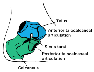
<span class="mw-page-title-main">Calcaneus</span> Bone of the tarsus of the foot

In humans and many other primates, the calcaneus or heel bone is a bone of the tarsus of the foot which constitutes the heel. In some other animals, it is the point of the hock.

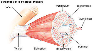
<span class="mw-page-title-main">Epimysium</span> Fibrous tissue envelope that surrounds skeletal muscle

Epimysium is the fibrous tissue envelope that surrounds skeletal muscle. It is a layer of dense irregular connective tissue which ensheaths the entire muscle and protects muscles from friction against other muscles and bones. It also allows a muscle to contract and move powerfully while maintaining its structural integrity.

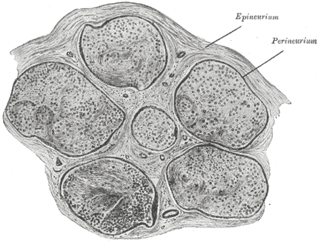
<span class="mw-page-title-main">Epineurium</span> Outermost layer of a nerve

The epineurium is the outermost layer of dense irregular connective tissue surrounding a peripheral nerve. It usually surrounds multiple nerve fascicles as well as blood vessels which supply the nerve. Smaller branches of these blood vessels penetrate into the perineurium. In addition to blood vessels which supply the nerve, lymphocytes and fibroblasts are also present and contribute to the production of collagen fibers that form the backbone of the epineurium. In addition to providing structural support, lymphocytes and fibroblasts also play a vital role in maintenance and repair of the surrounding tissues.

<span class="mw-page-title-main">Sagittal plane</span> Anatomical plane dividing the body into left and right

The sagittal plane is an anatomical plane that divides the body into right and left sections. It is perpendicular to the transverse and coronal planes. The plane may be in the center of the body and divide it into two equal parts (mid-sagittal), or away from the midline and divide it into unequal parts (para-sagittal).

<span class="mw-page-title-main">Occipitofrontalis muscle</span> Facial muscle helping to create facial expressions

The occipitofrontalis muscle is a muscle which covers parts of the skull. It consists of two parts or bellies: the occipital belly, near the occipital bone, and the frontal belly, near the frontal bone. It is supplied by the supraorbital artery, the supratrochlear artery, and the occipital artery. It is innervated by the facial nerve. In humans, the occipitofrontalis helps to create facial expressions.

<span class="mw-page-title-main">Infraorbital foramen</span> Opening in the maxillary bone of the skull

In human anatomy, the infraorbital foramen is one of two small holes in the skull's upper jawbone, located below the eye socket and to the left and right of the nose. Both holes are used for blood vessels and nerves. In anatomical terms, it is located below the infraorbital margin of the orbit. It transmits the infraorbital artery and vein, and the infraorbital nerve, a branch of the maxillary nerve. It is typically 6.10 to 10.9 mm from the infraorbital margin.

<span class="mw-page-title-main">Sympathetic trunk</span> Bundle of nerve fibers

The sympathetic trunks are a paired bundle of nerve fibers that run from the base of the skull to the coccyx. They are a major component of the sympathetic nervous system.

<span class="mw-page-title-main">Perineurium</span> Sheath around nerve fasciculi

The perineurium is a protective sheath that surrounds a nerve fascicle. This bundles together axons targeting the same anatomical location. The perineurium is composed from fibroblasts.

Matthew H. Kaufman was a British biologist. He was Professor Emeritus at University of Edinburgh having been Professor of Anatomy there from 1985 to 2007. He taught anatomy and embryology for more than 30 years, initially at the University of Cambridge, when he was a Fellow of King's College, and more recently in Edinburgh.

<span class="mw-page-title-main">Erection</span> Physiological phenomenon involving the hardening and enlargement of the penis

An erection is a physiological phenomenon in which the penis becomes firm, engorged, and enlarged. Penile erection is the result of a complex interaction of psychological, neural, vascular, and endocrine factors, and is often associated with sexual arousal, sexual attraction or libido, although erections can also be spontaneous. The shape, angle, and direction of an erection vary considerably between humans.

<span class="mw-page-title-main">Douglas DF</span> 1936 airliner flying boat series by Douglas

The Douglas DF was a commercial flying boat built by Douglas Aircraft Company, first flown on 24 September 1936.

<span class="mw-page-title-main">Vertebra</span> Bone in the vertebral column

Each vertebra is an irregular bone with a complex structure composed of bone and some hyaline cartilage, that make up the vertebral column or spine, of vertebrates. The proportions of the vertebrae differ according to their spinal segment and the particular species.

References

    1. "Cranial Nerves - Function, Table, Anatomy and FAQs".
    2. Butler, Ann B.; Hodos, William (2005). Comparative Vertebrate Neuroanatomy: Evolution and Adaptation. John Wiley & Sons. p. 605. ISBN   978-0-471-73383-6.
    3. McCracken, Thomas (2000). New Atlas of Human Anatomy. China: MetroBooks. pp. 1–240. ISBN   1-58663-097-0.
    4. McCracken, Thomass (2000). New Atlas of Human Anatomy. China: MetroBooks. pp. 1–240. ISBN   1-58663-097-0.