Magnocellular red nucleus

Last updated
Magnocellular red nucleus
Identifiers
NeuroLex ID birnlex_720
Anatomical terms of neuroanatomy

The magnocellular red nucleus (mRN or mNR or RNm) is located in the rostral midbrain and is involved in motor coordination. [1] [2] Together with the parvocellular red nucleus, the mRN makes up the red nucleus. [2] Due to the role it plays in motor coordination, the magnocellular red nucleus may be implicated in the characteristic symptom of restless legs syndrome (RLS). [3] The mRN receives most of its signals from the motor cortex and the cerebellum. [4] [5]

Contents

Overview

Section, in the transverse plane through the human midbrain at the level of the superior colliculus, that shows the rough path of each nervus oculomotorius from its nucleus toward the eye and the relative location of the red nucleus. For reference, the bottom of the picture is where the front of the head is located. Cn3nucleus-en.svg
Section, in the transverse plane through the human midbrain at the level of the superior colliculus, that shows the rough path of each nervus oculomotorius from its nucleus toward the eye and the relative location of the red nucleus. For reference, the bottom of the picture is where the front of the head is located.

The red nucleus (RN), a group of neurons composed of the parvocellular red nucleus (pRN) and the magnocellular red nucleus (mRN), contributes to movement and motor control within the forelimb. [6] Primate studies have shown that more forelimb mRN neuron discharges are observed when the location of the target object a primate is reaching is on the right or above. This demonstrates that although forelimb mRN neurons are involved in grasping movements to the left, right, above, and below, they play a greater role when an organism is attempting to reach an object to the right or above. [7] Scientists were also able to find differences in the functions and anatomies between the parvicellular and magnocellular regions of red nuclei in monkeys. Single unit recording in two monkeys that were kept awake during experimentation was used to search for functional differences between the two regions of the red nucleus. In order to investigate inputs to the two regions, anatomical tracing of WGA-HRP was used. Overall, the magnocellular region was much more responsive than that parvicellular region. [8] The pRN is located in the diencephalon, a division of the forebrain, and the mRN is located in the mesencephalon (midbrain). [6] Unlike the pRN, which is both GABAergic and glutamatergic, the mRN is solely glutamatergic. [9]

The RN is capable of projection onto the spinal cord interneurons and motor neurons of signals integrated from the motor cortex and cerebellum. [6] The mRN already existed in tetrapods after they became terrestrial. It became further developed in mammals when the volume of the mRN increased along with the development of the intermediate cerebellum and the interposed nuclei. [5]

Neuron Counts in pRN and mRN

The absolute number of pRN and mRN neurons in the midbrain can be estimated. An analysis of complete sets of serial 40-μm glycolmethacrylate sections can be used to find this number. This was done on six young adult male rats, and a complete set of these serials sections for each rat were used to quantify the neuronal numbers. [10] By using Cavalieri's method, the total volume of the red nucleus can be estimated. The optical dissector method on the same set of sampled sections is then used to estimate the numerical density Nv, or the number of neurons within a certain volume ("subvolume") of the nucleus. The absolute number of neurons can then be found by multiplying the total volume of the nucleus by the numerical density. In rat brains, there is an average of 8394 pRN and 6986 mRN. [10]

The number of pRN and mRN can be used to gain a better understanding of how organisms differ in motor abilities. It has been reported that mice have a total of around 3200 neurons in the red nucleus, while rats have an average of nearly 15,400 neurons. Compared to rats, mice have inferior motor abilities in skilled reaching tasks, which has been reported to potentially be a result of the varying neuron counts in the red nucleus. [11]

Development

Phylogenetically

The RN, consisting of the pRN and the mRN, can be found in land vertebrates and some species of rays. It is defined by its relative position and relation to its contralateral rubrospinal projections, tegmentum mesencephalic and crossed rubrospinal tract. Although a crossed rubrospinal tract is absent in boidae, sharks, and limbless amphibians, it is present in certain rays. The rays which have this use their pectoral fins to move. A version of the rubrospinal tract is found in lungfishes but not in advanced snakes, suggesting that the rubrospinal tract is directly related to the presence of limbs. The connectivity of the RN is determined by the type and complexity of motor function that land vertebrates are capable of. [12]

Ontogenetically

Separated immature RNm cells first appear around 12 weeks of gestation. They show up in clumps dorsal to the parvocellular red nucleus (RNp). The RNm then takes the shape of a crescent moon ventral to the RNp consisting of basophilic neurons. 18 to 23 weeks into gestation, the neurons then appear dorsal to the RNp, as island clusters among the myelinated oculomotor nerve roots. Around 28 weeks, the nerves are distributed ventrolateral to the superior cerebellar peduncle as well as around the caudal pole of the RNp. As gestation progresses, the neurons increase in volume, fracturing into two distinct sizes, which become clear at 33 weeks. In summary, the differentiation and maturation of the RNm slowly progresses in the later half of gestation, and the locations of the magnocellular red nuclei vary across the brain. [2] A 2012 study shows that the human fetus has a more developed mRN than an adult, because of its greater number of "myelinated fibers in the rubrospinal tracts." This suggests that the mRN plays a greater role earlier on in development than later on in life. [13]

Related Research Articles

<span class="mw-page-title-main">Hypothalamus</span> Area of the brain below the thalamus

The hypothalamus is a small part of the brain that contains a number of nuclei with a variety of functions. One of the most important functions is to link the nervous system to the endocrine system via the pituitary gland. The hypothalamus is located below the thalamus and is part of the limbic system. It forms the ventral part of the diencephalon. All vertebrate brains contain a hypothalamus. In humans, it is the size of an almond.

<span class="mw-page-title-main">Brainstem</span> Posterior part of the brain, adjoining and structurally continuous

The brainstem is the stalk-like part of the brain that interconnects the cerebrum and diencephalon with the spinal cord. In the human brain, the brainstem is composed of the midbrain, the pons, and the medulla oblongata. The midbrain is continuous with the thalamus of the diencephalon through the tentorial notch.

<span class="mw-page-title-main">Lateral geniculate nucleus</span> Component of the visual system in the brains thalamus

In neuroanatomy, the lateral geniculate nucleus is a structure in the thalamus and a key component of the mammalian visual pathway. It is a small, ovoid, ventral projection of the thalamus where the thalamus connects with the optic nerve. There are two LGNs, one on the left and another on the right side of the thalamus. In humans, both LGNs have six layers of neurons alternating with optic fibers.

<span class="mw-page-title-main">Extrapyramidal system</span> Connection between brain and spinal cord

In anatomy, the extrapyramidal system is a part of the motor system network causing involuntary actions. The system is called extrapyramidal to distinguish it from the tracts of the motor cortex that reach their targets by traveling through the pyramids of the medulla. The pyramidal tracts may directly innervate motor neurons of the spinal cord or brainstem, whereas the extrapyramidal system centers on the modulation and regulation of anterior (ventral) horn cells.

<span class="mw-page-title-main">Midbrain</span> Forward-most portion of the brainstem

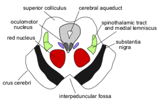
The midbrain or mesencephalon is the rostral-most portion of the brainstem connecting the diencephalon and cerebrum with the pons. It consists of the cerebral peduncles, tegmentum, and tectum.

<span class="mw-page-title-main">Paraventricular nucleus of hypothalamus</span>

The paraventricular nucleus is a nucleus in the hypothalamus. Anatomically, it is adjacent to the third ventricle and many of its neurons project to the posterior pituitary. These projecting neurons secrete oxytocin and a smaller amount of vasopressin, otherwise the nucleus also secretes corticotropin-releasing hormone (CRH) and thyrotropin-releasing hormone (TRH). CRH and TRH are secreted into the hypophyseal portal system and act on different targets neurons in the anterior pituitary. PVN is thought to mediate many diverse functions through these different hormones, including osmoregulation, appetite, and the response of the body to stress.

Magnocellular neurosecretory cells are large neuroendocrine cells within the supraoptic nucleus and paraventricular nucleus of the hypothalamus. They are also found in smaller numbers in accessory cell groups between these two nuclei, the largest one being the circular nucleus. There are two types of magnocellular neurosecretory cells, oxytocin-producing cells and vasopressin-producing cells, but a small number can produce both hormones. These cells are neuroendocrine neurons, are electrically excitable, and generate action potentials in response to afferent stimulation. Vasopressin is produced from the vasopressin-producing cells via the AVP gene, a molecular output of circadian pathways.

<span class="mw-page-title-main">Pretectal area</span> Structure in the midbrain which mediates responses to ambient light

In neuroanatomy, the pretectal area, or pretectum, is a midbrain structure composed of seven nuclei and comprises part of the subcortical visual system. Through reciprocal bilateral projections from the retina, it is involved primarily in mediating behavioral responses to acute changes in ambient light such as the pupillary light reflex, the optokinetic reflex, and temporary changes to the circadian rhythm. In addition to the pretectum's role in the visual system, the anterior pretectal nucleus has been found to mediate somatosensory and nociceptive information.

<span class="mw-page-title-main">Reticular formation</span> Spinal trigeminal nucleus

The reticular formation is a set of interconnected nuclei that are located in the brainstem, hypothalamus, and other regions. It is not anatomically well defined, because it includes neurons located in different parts of the brain. The neurons of the reticular formation make up a complex set of networks in the core of the brainstem that extend from the upper part of the midbrain to the lower part of the medulla oblongata. The reticular formation includes ascending pathways to the cortex in the ascending reticular activating system (ARAS) and descending pathways to the spinal cord via the reticulospinal tracts.

<span class="mw-page-title-main">Rubrospinal tract</span> Part of the nervous system

The rubrospinal tract is a part of the nervous system. It is a part of the lateral indirect extra-pyramidal tract.

<span class="mw-page-title-main">Red nucleus</span> Structure in the human brain

The red nucleus or nucleus ruber is a structure in the rostral midbrain involved in motor coordination. The red nucleus is pale pink, which is believed to be due to the presence of iron in at least two different forms: hemoglobin and ferritin. The structure is located in the tegmentum of the midbrain next to the substantia nigra and comprises caudal magnocellular and rostral parvocellular components. The red nucleus and substantia nigra are subcortical centers of the extrapyramidal motor system.

<span class="mw-page-title-main">Globose nucleus</span> Part of the interposed nucleus, a structure in the cerebellum.

The globose nucleus is one of the deep cerebellar nuclei. It is located medial to the emboliform nucleus, and lateral to the fastigial nucleus. The globose nucleus and emboliform nucleus are known collectively as the interposed nuclei.

The zona incerta (ZI) is a horizontally elongated region of gray matter in the subthalamus below the thalamus. Its connections project extensively over the brain from the cerebral cortex down into the spinal cord.

The rubro-olivary tract is a tract which connects the inferior olive and the parvocellular red nucleus.

<span class="mw-page-title-main">Central tegmental tract</span> Structure in the midbrain and pons

The central tegmental tract is a structure in the midbrain and pons.

<span class="mw-page-title-main">Anatomy of the cerebellum</span> Structures in the cerebellum, a part of the brain

The anatomy of the cerebellum can be viewed at three levels. At the level of gross anatomy, the cerebellum consists of a tightly folded and crumpled layer of cortex, with white matter underneath, several deep nuclei embedded in the white matter, and a fluid-filled ventricle in the middle. At the intermediate level, the cerebellum and its auxiliary structures can be broken down into several hundred or thousand independently functioning modules or compartments known as microzones. At the microscopic level, each module consists of the same small set of neuronal elements, laid out with a highly stereotyped geometry.

The parvocellular red nucleus (RNp) is located in the rostral midbrain and is involved in motor coordination. Together with the magnocellular red nucleus, it makes up the red nucleus.

Parvocellular neurosecretory cells are small neurons that produce hypothalamic releasing and inhibiting hormones. The cell bodies of these neurons are located in various nuclei of the hypothalamus or in closely related areas of the basal brain, mainly in the medial zone of the hypothalamus. All or most of the axons of the parvocellular neurosecretory cells project to the median eminence, at the base of the brain, where their nerve terminals release the hypothalamic hormones. These hormones are then immediately absorbed into the blood vessels of the hypothalamo-pituitary portal system, which carry them to the anterior pituitary gland, where they regulate the secretion of hormones into the systemic circulation.

The parabrachial nuclei, also known as the parabrachial complex, are a group of nuclei in the dorsolateral pons that surrounds the superior cerebellar peduncle as it enters the brainstem from the cerebellum. They are named from the Latin term for the superior cerebellar peduncle, the brachium conjunctivum. In the human brain, the expansion of the superior cerebellar peduncle expands the parabrachial nuclei, which form a thin strip of grey matter over most of the peduncle. The parabrachial nuclei are typically divided along the lines suggested by Baxter and Olszewski in humans, into a medial parabrachial nucleus and lateral parabrachial nucleus. These have in turn been subdivided into a dozen subnuclei: the superior, dorsal, ventral, internal, external and extreme lateral subnuclei; the lateral crescent and subparabrachial nucleus along the ventrolateral margin of the lateral parabrachial complex; and the medial and external medial subnuclei

<span class="mw-page-title-main">Mesencephalic locomotor region</span>

The mesencephalic locomotor region (MLR) is a functionally defined area of the midbrain that is associated with the initiation and control of locomotor movements in vertebrate species.

References

  1. Purves, Dale; Augustine, George C.; Fitzpatrick, David; Hall, William C.; LaMantia, Anthony-Samuel; White, Leonard E. (2012). Neuroscience (5th ed.). Sinauer Associates, Inc. pp. 269, 271–272. ISBN   978-0-87893-695-3.
  2. 1 2 3 Yamaguchi K, Goto N (August 2006). "Development of the human magnocellular red nucleus: a morphological study". Brain Dev. 28 (7): 431–5. doi:10.1016/j.braindev.2006.01.001. PMID   16516425. S2CID   8483275.
  3. Baier, P. C.; Trenkwalder, CLAUDIA (2005-01-01), LeDoux, Mark (ed.), "CHAPTER N1 - Clinical Features and Animal Models of Restless Legs Syndrome and Periodic Limb Movement" , Animal Models of Movement Disorders, Academic Press, pp. 755–758, doi:10.1016/b978-012088382-0/50067-0, ISBN   9780120883820 , retrieved 2019-11-02
  4. Gibson, A R; Houk, J C; Kohlerman, N J (January 1985). "Magnocellular red nucleus activity during different types of limb movement in the macaque monkey". The Journal of Physiology. 358: 527–549. doi:10.1113/jphysiol.1985.sp015565. ISSN   0022-3751. PMC   1193356 . PMID   3981472.
  5. 1 2 Padel, Y. (1993). "[Magnocellular and parvocellular red nuclei. Anatomico-functional aspects and relations with the cerebellum and other nerve centres]". Revue Neurologique. 149 (11): 703–715. ISSN   0035-3787. PMID   8091083.
  6. 1 2 3 Martinez-Lopez, Jesus E.; Moreno-Bravo, Juan A.; Madrigal, M. Pilar; Martinez, Salvador; Puelles, Eduardo (2015). "Red nucleus and rubrospinal tract disorganization in the absence of Pou4f1". Frontiers in Neuroanatomy. 9: 8. doi: 10.3389/fnana.2015.00008 . ISSN   1662-5129. PMC   4318420 . PMID   25698939.
  7. van Kan, Peter; McCurdy, Martha (2002-01-01). "Discharge of primate magnocellular red nucleus neurons during reaching to grasp in different spatial locations". Experimental Brain Research. 142 (1): 151–157. doi:10.1007/s00221-001-0924-5. ISSN   0014-4819. PMID   11797092. S2CID   42113292.
  8. Kennedy, P. R.; Gibson, A. R.; Houk, J. C. (1986-01-29). "Functional and anatomic differentiation between parvicellular and magnocellular regions of red nucleus in the monkey". Brain Research. 364 (1): 124–136. doi:10.1016/0006-8993(86)90993-5. ISSN   0006-8993. PMID   3947959. S2CID   32770040.
  9. Watson, Charles, 1943- Paxinos, George, 1944- Puelles, L. (Luis) (2012). The mouse nervous system. Elsevier Academic Press. ISBN   9780080919188. OCLC   760157364.{{cite book}}: CS1 maint: multiple names: authors list (link) CS1 maint: numeric names: authors list (link)
  10. 1 2 Aghoghovwia, Benjamin E.; Oorschot, Dorothy E. (September 2016). "Absolute number of parvicellular and magnocellular neurons in the red nucleus of the rat midbrain: a stereological study". Journal of Anatomy. 229 (3): 406–415. doi:10.1111/joa.12495. ISSN   0021-8782. PMC   4974543 . PMID   27257130.
  11. Whishaw, I. Q. (August 1996). "An endpoint, descriptive, and kinematic comparison of skilled reaching in mice (Mus musculus) with rats (Rattus norvegicus)". Behavioural Brain Research. 78 (2): 101–111. doi:10.1016/0166-4328(95)00236-7. ISSN   0166-4328. PMID   8864042. S2CID   27333748.
  12. ten Donkelaar, H. J. (April 1988). "Evolution of the red nucleus and rubrospinal tract". Behavioural Brain Research. 28 (1–2): 9–20. doi:10.1016/0166-4328(88)90072-1. ISSN   0166-4328. PMID   3289562. S2CID   54367413.
  13. Hicks, T. Philip; Onodera, Satoru (2012-02-01). "The mammalian red nucleus and its role in motor systems, including the emergence of bipedalism and language". Progress in Neurobiology. 96 (2): 165–175. doi:10.1016/j.pneurobio.2011.12.002. ISSN   0301-0082. PMID   22230734. S2CID   22481162.