Fibrinolysis

Last updated

Fibrinolysis is a process that prevents blood clots from growing and becoming problematic. [1] Primary fibrinolysis is a normal body process, while secondary fibrinolysis is the breakdown of clots due to a medicine, a medical disorder, or some other cause. [2]

Contents

In fibrinolysis, a fibrin clot, the product of coagulation, is broken down. [3] Its main enzyme plasmin cuts the fibrin mesh at various places, leading to the production of circulating fragments that are cleared by other proteases or by the kidney and liver.

Physiology

Fibrinolysis (simplified). Blue arrows denote stimulation, and red arrows inhibition. Fibrinolysis.svg
Fibrinolysis (simplified). Blue arrows denote stimulation, and red arrows inhibition.

Plasmin is produced in an inactive form, plasminogen, in the liver. Although plasminogen cannot cleave fibrin, it still has an affinity for it, and is incorporated into the clot when it is formed.

Tissue plasminogen activator (t-PA) [4] and urokinase are the agents that convert plasminogen to the active plasmin, thus allowing fibrinolysis to occur. t-PA is released into the blood slowly by the damaged endothelium of the blood vessels, such that, after several days (when the bleeding has stopped), the clot is broken down. This occurs because plasminogen became entrapped within the clot when it formed; as it is slowly activated, it breaks down the fibrin mesh. t-PA and urokinase are themselves inhibited by plasminogen activator inhibitor-1 and plasminogen activator inhibitor-2 (PAI-1 and PAI-2). In contrast, plasminogen further stimulates plasmin generation by producing more active forms of both tissue plasminogen activator (tPA) and urokinase.

α2-Antiplasmin and α2-macroglobulin inactivate plasmin. Plasmin activity is also reduced by thrombin-activatable fibrinolysis inhibitor (TAFI), which modifies fibrin to make it more resistant to the tPA-mediated plasminogen.

Measurement

Plasmin breaks down fibrin into soluble parts called fibrin degradation products (FDPs). FDPs compete with thrombin, and thus slow down clot formation by preventing the conversion of fibrinogen to fibrin. This effect can be seen in the thrombin clotting time (TCT) test, which is prolonged in a person that has active fibrinolysis.

Antibody-antigen technology can measure FDPs and a specific FDP, the D-dimer. This is more specific than the TCT, and confirms that fibrinolysis has occurred. It is therefore used to indicate deep-vein thrombosis, pulmonary embolism, DIC, and efficacy of treatment in acute myocardial infarction. Alternatively, a more rapid detection of fibrinolytic activity, especially hyperfibrinolysis, is possible with thromboelastometry (TEM) in whole blood, even in patients on heparin. In this assay, increased fibrinolysis is assessed by comparing the TEM profile in the absence or presence of the fibrinolysis inhibitor aprotinin. Clinically, the TEM is useful for near real-time measurement of activated fibrinolysis for at-risk patients, such as those experiencing significant blood loss during surgery. [5]

Testing of overall fibrinolysis can be measured by a euglobulin lysis time (ELT) assay. The ELT measures fibrinolysis by clotting the euglobulin fraction (primarily the fibrinolytic factors fibrinogen, PAI-1, tPA, α2-antiplasmin, and plasminogen) from plasma and then observing the time required for clot dissolution. A shortened lysis time indicates a hyperfibrinolytic state and bleeding risk. Such results can be seen in peoples with liver disease, PAI-1 deficiency or α2-antiplasmin deficiency. Similar results are also seen after administration of desmopressin or after severe stress. [6]

Role in disease

Few congenital disorders of the fibrinolytic system have been documented. Nevertheless, excess levels of PAI and α2-antiplasmin have been implicated in metabolic syndrome and various other disease states.

However, acquired disturbance of fibrinolysis (hyperfibrinolysis), is not uncommon. Many trauma patients have an overwhelming activation of tissue factor and thus massive hyperfibrinolysis. [7] Hyperfibrinolysis may occur in other disease states. It could lead to massive bleeding if not diagnosed and treated early enough.

The fibrinolytic system is closely linked to control of inflammation, and plays a role in disease states associated with inflammation. Plasmin, in addition to lysing fibrin clots, also cleaves the complement system component C3, and fibrin degradation products have some vascular permeability inducing effects.

Pharmacology

In a process called thrombolysis (the breakdown of a thrombus), fibrinolytic drugs are used. They are given following a heart attack to dissolve the thrombus blocking the coronary artery; experimentally after a stroke to allow blood flow back to the affected part of the brain; and in the event of pulmonary embolism. [8]

Thrombolysis refers to the dissolution of the thrombus due to various agents while fibrinolysis refers specifically to the agents causing fibrin breakdown in the clot.

Antifibrinolytics, such as aminocaproic acid (ε-aminocaproic acid) and tranexamic acid are used as inhibitors of fibrinolysis. Their application may be beneficial in patients with hyperfibrinolysis because they arrest bleeding rapidly if the other components of the haemostatic system are not severely affected. [9] This may help to avoid the use of blood products such as fresh frozen plasma with its associated risks of infections or anaphylactic reactions.

Fibrinolytic enzymes

Related Research Articles

<span class="mw-page-title-main">Thrombus</span> Blood clot

A thrombus, colloquially called a blood clot, is the final product of the blood coagulation step in hemostasis. There are two components to a thrombus: aggregated platelets and red blood cells that form a plug, and a mesh of cross-linked fibrin protein. The substance making up a thrombus is sometimes called cruor. A thrombus is a healthy response to injury intended to stop and prevent further bleeding, but can be harmful in thrombosis, when a clot obstructs blood flow through a healthy blood vessel in the circulatory system.

<span class="mw-page-title-main">Coagulation</span> Process of formation of blood clots

Coagulation, also known as clotting, is the process by which blood changes from a liquid to a gel, forming a blood clot. It results in hemostasis, the cessation of blood loss from a damaged vessel, followed by repair. The process of coagulation involves activation, adhesion and aggregation of platelets, as well as deposition and maturation of fibrin.

<span class="mw-page-title-main">Disseminated intravascular coagulation</span> Medical condition where blood clots block small blood vessels

Disseminated intravascular coagulation (DIC) is a condition in which blood clots form throughout the body, blocking small blood vessels. Symptoms may include chest pain, shortness of breath, leg pain, problems speaking, or problems moving parts of the body. As clotting factors and platelets are used up, bleeding may occur. This may include blood in the urine, blood in the stool, or bleeding into the skin. Complications may include organ failure.
<span class="mw-page-title-main">Fibrinogen</span> Soluble protein complex in blood plasma and involved in clot formation

Fibrinogen is a glycoprotein complex, produced in the liver, that circulates in the blood of all vertebrates. During tissue and vascular injury, it is converted enzymatically by thrombin to fibrin and then to a fibrin-based blood clot. Fibrin clots function primarily to occlude blood vessels to stop bleeding. Fibrin also binds and reduces the activity of thrombin. This activity, sometimes referred to as antithrombin I, limits clotting. Fibrin also mediates blood platelet and endothelial cell spreading, tissue fibroblast proliferation, capillary tube formation, and angiogenesis and thereby promotes revascularization and wound healing.

<span class="mw-page-title-main">Thrombolysis</span> Breakdown (lysis) of blood clots formed in blood vessels, using medication

Thrombolysis, also called fibrinolytic therapy, is the breakdown (lysis) of blood clots formed in blood vessels, using medication. It is used in ST elevation myocardial infarction, stroke, and in cases of severe venous thromboembolism.
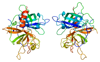
<span class="mw-page-title-main">Tissue-type plasminogen activator</span> Protein involved in the breakdown of blood clots

Tissue-type plasminogen activator, short name tPA, is a protein that facilitates the breakdown of blood clots. It acts as an enzyme to convert plasminogen into its active form plasmin, the major enzyme responsible for clot breakdown. It is a serine protease found on endothelial cells lining the blood vessels. Human tPA is encoded by the PLAT gene, and has a molecular weight of ~70 kDa in the single-chain form.

<span class="mw-page-title-main">Plasmin</span> Enzyme in human blood that degrades clots and other proteins

Plasmin is an important enzyme present in blood that degrades many blood plasma proteins, including fibrin clots. The degradation of fibrin is termed fibrinolysis. In humans, the plasmin protein is encoded by the PLG gene.

<span class="mw-page-title-main">Urokinase</span> Human protein

Urokinase, also known as urokinase-type plasminogen activator (uPA), is a serine protease present in humans and other animals. The human urokinase protein was discovered, but not named, by McFarlane and Pilling in 1947. Urokinase was originally isolated from human urine, and it is also present in the blood and in the extracellular matrix of many tissues. The primary physiological substrate of this enzyme is plasminogen, which is an inactive form (zymogen) of the serine protease plasmin. Activation of plasmin triggers a proteolytic cascade that, depending on the physiological environment, participates in thrombolysis or extracellular matrix degradation. This cascade had been involved in vascular diseases and cancer progression.
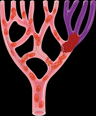
<span class="mw-page-title-main">Alteplase</span> Thrombolytic medication

Alteplase, sold under the brand name Activase among others, is a biosynthetic form of human tissue-type plasminogen activator (t-PA). It is a thrombolytic medication used to treat acute ischemic stroke, acute ST-elevation myocardial infarction, pulmonary embolism associated with low blood pressure, and blocked central venous catheter. Alteplase is given by injection into a vein or artery. Alteplase is the same as the normal human plasminogen activator produced in vascular endothelial cells and is synthesized via recombinant DNA technology in Chinese hamster ovary cells (CHO). Alteplase causes the breakdown of a clot by inducing fibrinolysis.

<span class="mw-page-title-main">Alpha 2-antiplasmin</span> Protein-coding gene in the species Homo sapiens

Alpha 2-antiplasmin is a serine protease inhibitor (serpin) responsible for inactivating plasmin. Plasmin is an important enzyme that participates in fibrinolysis and degradation of various other proteins. This protein is encoded by the SERPINF2 gene.
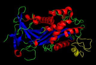
<span class="mw-page-title-main">Plasminogen activator inhibitor-1</span> Human protein

Plasminogen activator inhibitor-1 (PAI-1) also known as endothelial plasminogen activator inhibitor is a protein that in humans is encoded by the SERPINE1 gene. Elevated PAI-1 is a risk factor for thrombosis and atherosclerosis.

<span class="mw-page-title-main">Plasminogen activator</span> Type of protein

Plasminogen activators are serine proteases that catalyze the activation of plasmin via proteolytic cleavage of its zymogen form plasminogen. Plasmin is an important factor in fibrinolysis, the breakdown of fibrin polymers formed during blood clotting. There are two main plasminogen activators: urokinase (uPA) and tissue plasminogen activator (tPA). Tissue plasminogen activators are used to treat medical conditions related to blood clotting including embolic or thrombotic stroke, myocardial infarction, and pulmonary embolism.

<span class="mw-page-title-main">Aminocaproic acid</span> Chemical compound

Aminocaproic acid is a derivative and analogue of the amino acid lysine, which makes it an effective inhibitor for enzymes that bind that particular residue. Such enzymes include proteolytic enzymes like plasmin, the enzyme responsible for fibrinolysis. For this reason it is effective in treatment of certain bleeding disorders, and it is sold under the brand name Amicar. Aminocaproic acid is also an intermediate in the polymerization of Nylon-6, where it is formed by ring-opening hydrolysis of caprolactam. The crystal structure determination showed that the 6-aminohexanoic acid is present as a salt, at least in the solid state.

Antifibrinolytics are a class of medication that are inhibitors of fibrinolysis. Examples include aminocaproic acid and tranexamic acid. These lysine-like drugs interfere with the formation of the fibrinolytic enzyme plasmin from its precursor plasminogen by plasminogen activators which takes place mainly in lysine rich areas on the surface of fibrin.

Tenecteplase, sold under the trade names TNKase, Metalyse and Elaxim, is an enzyme used as a thrombolytic drug.

<span class="mw-page-title-main">Désiré Collen</span> Belgian chemist, physician

Désiré, Baron Collen is a Belgian physician, chemist, biotechnology entrepreneur and life science investor. He made several discoveries in thrombosis, haemostasis and vascular biology in many of which serendipity played a significant role. His main achievement has been his role in the development of tissue-type plasminogen activator (t-PA) from a laboratory concept to a life-saving drug for dissolving blood clots causing acute myocardial infarction or acute ischemic stroke. Recombinant t-PA was produced and marketed by Genentech Inc as Activase and by Boehringer Ingelheim GmbH as Actilyse, and is considered biotechnology's first life saving drug.

<span class="mw-page-title-main">Quebec platelet disorder</span> Medical condition

Quebec platelet disorder (QPD) is a rare autosomal dominant bleeding disorder first described in a family from the province of Quebec, Canada. The disorder is characterized by large amounts of the fibrinolytic enzyme urokinase-type plasminogen activator (uPA) in platelets. This causes accelerated fibrinolysis which can result in bleeding.

The fibrinolysis system is responsible for removing blood clots. Hyperfibrinolysis describes a situation with markedly enhanced fibrinolytic activity, resulting in increased, sometimes catastrophic bleeding. Hyperfibrinolysis can be caused by acquired or congenital reasons. Among the congenital conditions for hyperfibrinolysis, alpha-2-plasmin inhibitor deficiency or plasminogen activator inhibitor type 1 (PAI-1) are very rare. The affected individuals show a hemophilia-like bleeding phenotype. Acquired hyperfibrinolysis is found in liver disease, in patients with severe trauma, during major surgical procedures, and other conditions. A special situation with temporarily enhanced fibrinolysis is thrombolytic therapy with drugs which activate plasminogen, e.g. for use in acute ischemic events or in patients with stroke. In patients with severe trauma, hyperfibrinolysis is associated with poor outcome. Moreover, hyperfibrinolysis may be associated with blood brain barrier impairment, a plasmin-dependent effect due to an increased generation of bradykinin.

Plasmin-α2-antiplasmin complex (PAP) is a 1:1 irreversibly formed inactive complex of the enzyme plasmin and its inhibitor α2-antiplasmin. It is a marker of the activity of the fibrinolytic system and a marker of net activation of fibrinolysis.

Alpha-2-plasmin inhibitor deficiency, also known as alpha-2-antiplasmindeficiency or congenital alpha-2-antiplasmin deficiency, is a rare autosomal recessive coagulopathy characterized by impaired inhibition of plasmin, leading to increased fibrinolysis and a heightened risk of bleeding.

References

  1. Mckenzie, Samuel (2019-03-27). "What is Fibrinolysis?". News-Medical. Retrieved 2024-05-31.
  2. Dugdale D. "Fibrinolysis - primary or secondary". MedlinePlus. Retrieved August 7, 2011.
  3. Cesarman-Maus G, Hajjar KA (May 2005). "Molecular mechanisms of fibrinolysis". British Journal of Haematology. 129 (3): 307–21. doi: 10.1111/j.1365-2141.2005.05444.x . PMID   15842654.
  4. Cotran RS, Kumar V, Collins T, Robbins SL (1999). Robbins pathologic basis of disease. Philadelphia: Saunders. ISBN   0-7216-7335-X. OCLC   39465455.
  5. Levrat A, Gros A, Rugeri L, Inaba K, Floccard B, Negrier C, David JS (June 2008). "Evaluation of rotation thrombelastography for the diagnosis of hyperfibrinolysis in trauma patients". Br J Anaesth. 100 (6): 792–7. doi: 10.1093/bja/aen083 . PMID   18440953.}
  6. Goodnight SH, Hathaway WE (2001). Disorders of hemostasis and thrombosis: a clinical guide. New York: McGraw-Hill, Medical Pub. Division. ISBN   0-07-134834-4. OCLC   45485184.
  7. Tieu BH, Holcomb JB, Schreiber MA (May 2007). "Coagulopathy: its pathophysiology and treatment in the injured patient". World J Surg. 31 (5): 1055–64. doi:10.1007/s00268-006-0653-9. PMID   17426904.}
  8. Patel N, Patel NJ, Agnihotri K, Panaich SS, Thakkar B, Patel A, et al. (December 2015). "Utilization of catheter-directed thrombolysis in pulmonary embolism and outcome difference between systemic thrombolysis and catheter-directed thrombolysis". Catheter Cardiovasc Interv. 86 (7): 1219–27. doi: 10.1002/ccd.26108 . PMID   26308961.}
  9. Levy JH, Koster A, Quinones QJ, Milling TJ, Key NS (March 2018). "Antifibrinolytic Therapy and Perioperative Considerations". Anesthesiology. 128 (3): 657–670. doi:10.1097/ALN.0000000000001997. PMC   5811331 . PMID   29200009.}