Factor V

Last updated
F5
Protein F5 PDB 1czs.png
Available structures
PDB Ortholog search: PDBe RCSB
Identifiers
Aliases F5 , FVL, PCCF, RPRGL1, THPH2, coagulation factor V
External IDs OMIM: 612309 MGI: 88382 HomoloGene: 104 GeneCards: F5
Orthologs
SpeciesHumanMouse
Entrez
Ensembl
UniProt
RefSeq (mRNA)

NM_000130

NM_007976

RefSeq (protein)

NP_000121

NP_032002

Location (UCSC) Chr 1: 169.51 – 169.59 Mb Chr 1: 163.98 – 164.05 Mb
PubMed search [3] [4]
Wikidata
View/Edit Human View/Edit Mouse

Factor V (pronounced factor five) is a protein of the coagulation system, rarely referred to as proaccelerin or labile factor. In contrast to most other coagulation factors, it is not enzymatically active but functions as a cofactor. [5] Deficiency leads to predisposition for hemorrhage, while some mutations (most notably factor V Leiden) predispose for thrombosis.

Contents

Genetics

The gene for factor V is located on the first chromosome (1q24). It is genomically related to the family of multicopper oxidases, and is homologous to coagulation factor VIII. The gene spans 70 kb, consists of 25 exons, and the resulting protein has a relative molecular mass of approximately 330kDa.

Structure

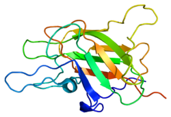
Factor V protein consists of six domains: A1-A2-B-A3-C1-C2.

The A domains are homologous to the A domains of the copper-binding protein ceruloplasmin, and form a triangular as in that protein. A copper ion is bound in the A1-A3 interface, and A3 interacts with the plasma. [6]

The C domains belong to the phospholipid-binding discoidin domain family (unrelated to C2 domain), and the C2 domain mediates membrane binding. The B domain C-terminus acts as a cofactor for the anticoagulant protein C activation by protein S. [7] [8]

Activation of factor V to factor Va is done by cleavage and release of the B domain, after which the protein no longer assists in activating protein C. The protein is now divided to a heavy chain, consisting of the A1-A2 domains, and a light chain, consisting of the A3-C1-C2 domains. Both form non-covalently a complex in a calcium-dependent manner. This complex is the pro-coagulant factor Va. [7]

Physiology

Factor V is produced by megakaryocytes, which produce platelets and platelet-derived factor V, and hepatocytes, which produce plasma-derived factor V. [9] The molecule circulates in plasma as a single-chain molecule with a plasma half-life of 12–36 hours. [10]

Factor V is able to bind to activated platelets and is activated by thrombin. On activation, factor V is spliced in two chains (heavy and light chain with molecular masses of 110000 and 73000, respectively) which are noncovalently bound to each other by calcium. The thereby activated factor V (now called FVa) is a cofactor of the prothrombinase complex: The activated factor X (FXa) enzyme requires calcium and activated factor V (FVa) to convert prothrombin to thrombin on the cell surface membrane.

Factor Va is degraded by activated protein C, one of the principal physiological inhibitors of coagulation. In the presence of thrombomodulin, thrombin acts to decrease clotting by activating protein C; therefore, the concentration and action of protein C are important determinants in the negative feedback loop through which thrombin limits its own activation.

Role in disease

Various hereditary disorders of factor V are known. Deficiency is associated with a rare mild form of hemophilia (termed parahemophilia or Owren parahemophilia), the incidence of which is about 1:1,000,000. It inherits in an autosomal recessive fashion.

There exists a bleeding tendency associated with the genetic up‐regulation of FV‐short, a minor splicing isoform of FV. This abnormal bleeding tendency occurs in east Texas bleeding disorder, Amsterdam bleeding disorder, and a third and more extreme example described in 2021 by Karen L. Zimowski et al. [11]

Other mutations of factor V are associated with venous thrombosis. They are the most common hereditary causes for thrombophilia (a tendency to form blood clots). The most common one of these, factor V Leiden, is due to the replacement of an arginine residue with glutamine at amino acid position 506 (R506Q). All prothrombotic factor V mutations (factor V Leiden, factor V Cambridge, factor V Hong Kong) make it resistant to cleavage by activated protein C ("APC resistance"). It therefore remains active and increases the rate of thrombin generation.

History

Until the discovery of factor V, coagulation was regarded as a product of four factors: calcium (IV) and thrombokinase (III) together acting on prothrombin (II) to produce fibrinogen (I); this model had been outlined by Paul Morawitz in 1905. [12]

The suggestion that an additional factor might exist was made by Paul Owren  [ no ] (1905–1990), a Norwegian physician, during his investigations into the bleeding tendency of a lady called Mary (1914–2002). She had suffered from nosebleeds and menorrhagia (excessive menstrual blood loss) for most her life, and was found to have a prolonged prothrombin time, suggesting either vitamin K deficiency or chronic liver disease leading to prothrombin deficiency. However, neither were the case, and Owren demonstrated this by correcting the abnormality with plasma from which prothrombin had been removed. Using Mary's serum as index, he found that the "missing" factor, which he labeled V (I–IV having been used in Morawitz' model), had particular characteristics. Most investigations were performed during the Second World War, and while Owren published his results in Norway in 1944, he could not publish them internationally until the war was over. They appeared finally in The Lancet in 1947. [12] [13]

The possibility of an extra coagulation factor was initially resisted on methodological grounds by Drs Armand Quick and Walter Seegers, both world authorities in coagulation. Confirmatory studies from other groups led to their final approval several years later. [12]

Owren initially felt that factor V (labile factor or proaccelerin) activated another factor, which he named VI. VI was the factor that accelerated the conversion from prothrombin to thrombin. It was later discovered that factor V was "converted" (activated) by thrombin itself, and later still that factor VI was simply the activated form of factor V. [12]

The complete amino acid sequence of the protein was published in 1987. [14] In 1994 factor V Leiden, resistant to inactivation by protein C, was described; this abnormality is the most common genetic cause for thrombosis. [15]

Interactions

Factor V has been shown to interact with Protein S. [16] [17]

Related Research Articles

<span class="mw-page-title-main">Coagulation</span> Process of formation of blood clots

Coagulation, also known as clotting, is the process by which blood changes from a liquid to a gel, forming a blood clot. It potentially results in hemostasis, the cessation of blood loss from a damaged vessel, followed by repair. The mechanism of coagulation involves activation, adhesion and aggregation of platelets, as well as deposition and maturation of fibrin.

Factor V Leiden is a variant of human factor V, which causes an increase in blood clotting (hypercoagulability). Due to this mutation, protein C, an anticoagulant protein that normally inhibits the pro-clotting activity of factor V, is not able to bind normally to factor V, leading to a hypercoagulable state, i.e., an increased tendency for the patient to form abnormal and potentially harmful blood clots. Factor V Leiden is the most common hereditary hypercoagulability disorder amongst ethnic Europeans. It is named after the Dutch city of Leiden, where it was first identified in 1994 by Rogier Maria Bertina under the direction of Pieter Hendrick Reitsma. Despite the increased risk of venous thromboembolisms, people with one copy of this gene have not been found to have shorter lives than the general population. It is an autosomal dominant genetic disorder with incomplete penetrance.

<span class="mw-page-title-main">Fibrinogen</span> Soluble protein complex in blood plasma and involved in clot formation

Fibrinogen is a glycoprotein complex, produced in the liver, that circulates in the blood of all vertebrates. During tissue and vascular injury, it is converted enzymatically by thrombin to fibrin and then to a fibrin-based blood clot. Fibrin clots function primarily to occlude blood vessels to stop bleeding. Fibrin also binds and reduces the activity of thrombin. This activity, sometimes referred to as antithrombin I, limits clotting. Fibrin also mediates blood platelet and endothelial cell spreading, tissue fibroblast proliferation, capillary tube formation, and angiogenesis and thereby promotes revascularization and wound healing.

<span class="mw-page-title-main">Thrombin</span> Enzyme involved in blood coagulation in humans

Thrombin is a serine protease, an enzyme that, in humans, is encoded by the F2 gene.

<span class="mw-page-title-main">Antithrombin</span> Mammalian protein found in Homo sapiens

Antithrombin (AT) is a small glycoprotein that inactivates several enzymes of the coagulation system. It is a 464-amino-acid protein produced by the liver. It contains three disulfide bonds and a total of four possible glycosylation sites. α-Antithrombin is the dominant form of antithrombin found in blood plasma and has an oligosaccharide occupying each of its four glycosylation sites. A single glycosylation site remains consistently un-occupied in the minor form of antithrombin, β-antithrombin. Its activity is increased manyfold by the anticoagulant drug heparin, which enhances the binding of antithrombin to factor IIa (thrombin) and factor Xa.

<span class="mw-page-title-main">Factor VIII</span> Blood-clotting protein

Factor VIII (FVIII) is an essential blood-clotting protein, also known as anti-hemophilic factor (AHF). In humans, factor VIII is encoded by the F8 gene. Defects in this gene result in hemophilia A, an X-linked coagulation disorder. Factor VIII is produced in the liver’s sinusoidal cells and endothelial cells outside the liver throughout the body. This protein circulates in the bloodstream in an inactive form, bound to another molecule called von Willebrand factor, until an injury that damages blood vessels occurs. In response to injury, coagulation factor VIII is activated and separates from von Willebrand factor. The active protein interacts with another coagulation factor called factor IX. This interaction sets off a chain of additional chemical reactions that form a blood clot.

von Willebrand factor Mammalian protein involved in blood clotting

Von Willebrand factor (VWF) is a blood glycoprotein that promotes hemostasis, specifically, platelet adhesion. It is deficient and/or defective in von Willebrand disease and is involved in many other diseases, including thrombotic thrombocytopenic purpura, Heyde's syndrome, and possibly hemolytic–uremic syndrome. Increased plasma levels in many cardiovascular, neoplastic, metabolic, and connective tissue diseases are presumed to arise from adverse changes to the endothelium, and may predict an increased risk of thrombosis.

<span class="mw-page-title-main">Partial thromboplastin time</span> Test for coagulation of blood

The partial thromboplastin time (PTT), also known as the activated partial thromboplastin time, is a blood test that characterizes coagulation of the blood. A historical name for this measure is the kaolin-cephalin clotting time (KCCT), reflecting kaolin and cephalin as materials historically used in the test. Apart from detecting abnormalities in blood clotting, partial thromboplastin time is also used to monitor the treatment effect of heparin, a widely prescribed drug that reduces blood's tendency to clot.

<span class="mw-page-title-main">Factor IX</span> Protein involved in blood clotting in humans

Factor IX is one of the serine proteases of the coagulation system; it belongs to peptidase family S1. Deficiency of this protein causes haemophilia B. It was discovered in 1952 after a young boy named Stephen Christmas was found to be lacking this exact factor, leading to haemophilia.

<span class="mw-page-title-main">Protein S deficiency</span> Medical condition

Protein S deficiency is a disorder associated with increased risk of venous thrombosis. Protein S, a vitamin K-dependent physiological anticoagulant, acts as a nonenzymatic cofactor to activate protein C in the degradation of factor Va and factor VIIIa.

<span class="mw-page-title-main">Protein S</span>

Protein S is a vitamin K-dependent plasma glycoprotein synthesized in the liver. In the circulation, Protein S exists in two forms: a free form and a complex form bound to complement protein C4b-binding protein (C4BP). In humans, protein S is encoded by the PROS1 gene. Protein S plays a role in coagulation.

<span class="mw-page-title-main">Protein C</span> Mammalian protein found in Homo sapiens

Protein C, also known as autoprothrombin IIA and blood coagulation factor XIV, is a zymogen, that is, an inactive enzyme. The activated form plays an important role in regulating anticoagulation, inflammation, and cell death and maintaining the permeability of blood vessel walls in humans and other animals. Activated protein C (APC) performs these operations primarily by proteolytically inactivating proteins Factor Va and Factor VIIIa. APC is classified as a serine protease since it contains a residue of serine in its active site. In humans, protein C is encoded by the PROC gene, which is found on chromosome 2.

<span class="mw-page-title-main">Thrombophilia</span> Abnormality of blood coagulation

Thrombophilia is an abnormality of blood coagulation that increases the risk of thrombosis. Such abnormalities can be identified in 50% of people who have an episode of thrombosis that was not provoked by other causes. A significant proportion of the population has a detectable thrombophilic abnormality, but most of these develop thrombosis only in the presence of an additional risk factor.

<span class="mw-page-title-main">Factor X</span> Mammalian protein found in Homo sapiens

Factor X, also known by the eponym Stuart–Prower factor, is an enzyme of the coagulation cascade. It is a serine endopeptidase. Factor X is synthesized in the liver and requires vitamin K for its synthesis.

<span class="mw-page-title-main">Factor XI</span> Mammalian protein found in Homo sapiens

Factor XI or plasma thromboplastin antecedent is the zymogen form of factor XIa, one of the enzymes of the coagulation cascade. Like many other coagulation factors, it is a serine protease. In humans, Factor XI is encoded by the F11 gene.

<span class="mw-page-title-main">Tissue factor</span> Protein involved in blood coagulation

Tissue factor, also called platelet tissue factor, factor III, or CD142, is a protein encoded by the F3 gene, present in subendothelial tissue and leukocytes. Its role in the clotting process is the initiation of thrombin formation from the zymogen prothrombin. Thromboplastin defines the cascade that leads to the activation of factor X—the tissue factor pathway. In doing so, it has replaced the previously named extrinsic pathway in order to eliminate ambiguity.

The prothrombinase enzyme complex consists of factor Xa (a serine protease) and factor Va (a protein cofactor). The complex assembles on negatively charged phospholipid membranes in the presence of calcium ions. The prothrombinase complex catalyzes the conversion of prothrombin (factor II), an inactive zymogen, to thrombin (factor IIa), an active serine protease. The activation of thrombin is a critical reaction in the coagulation cascade, which functions to regulate hemostasis in the body. To produce thrombin, the prothrombinase complex cleaves two peptide bonds in prothrombin, one after Arg271 and the other after Arg320. Although it has been shown that factor Xa can activate prothrombin when unassociated with the prothrombinase complex, the rate of thrombin formation is severely decreased under such circumstances. The prothrombinase complex can catalyze the activation of prothrombin at a rate 3 x 10⁵-fold faster than can factor Xa alone. Thus, the prothrombinase complex is required for the efficient production of activated thrombin and also for adequate hemostasis.

<span class="mw-page-title-main">Activated protein C resistance</span> Medical condition

Activated protein C resistance (APCR) is a hypercoagulability characterized by a lack of a response to activated protein C (APC), which normally helps prevent blood from clotting excessively. This results in an increased risk of venous thrombosis, which resulting in medical conditions such as deep vein thrombosis and pulmonary embolism. The most common cause of hereditary APC resistance is factor V Leiden mutation.

Blood clotting tests are the tests used for diagnostics of the hemostasis system. Coagulometer is the medical laboratory analyzer used for testing of the hemostasis system. Modern coagulometers realize different methods of activation and observation of development of blood clots in blood or in blood plasma.

Björn Dahlbäck is a Swedish physician, medical researcher, and professor of clinical chemistry, specializing in hematology and the molecular mechanisms of blood coagulation. He determined that activated protein C (APC) resistance is the most common inherited risk factor of venous thrombosis.

References

  1. 1 2 3 GRCh38: Ensembl release 89: ENSG00000198734 - Ensembl, May 2017
  2. 1 2 3 GRCm38: Ensembl release 89: ENSMUSG00000026579 - Ensembl, May 2017
  3. "Human PubMed Reference:". National Center for Biotechnology Information, U.S. National Library of Medicine.
  4. "Mouse PubMed Reference:". National Center for Biotechnology Information, U.S. National Library of Medicine.
  5. "F5 gene: MedlinePlus Genetics". medlineplus.gov. Retrieved 2023-03-25.
  6. Villoutreix BO, Dahlbäck B (June 1998). "Structural investigation of the A domains of human blood coagulation factor V by molecular modeling". Protein Science. 7 (6): 1317–25. doi:10.1002/pro.5560070607. PMC   2144041 . PMID   9655335.
  7. 1 2 Thorelli E, Kaufman RJ, Dahlbäck B (June 1998). "The C-terminal region of the factor V B-domain is crucial for the anticoagulant activity of factor V". The Journal of Biological Chemistry. 273 (26): 16140–45. doi: 10.1074/jbc.273.26.16140 . PMID   9632668.
  8. Macedo-Ribeiro S, Bode W, Huber R, Quinn-Allen MA, Kim SW, Ortel TL, Bourenkov GP, Bartunik HD, Stubbs MT, Kane WH, Fuentes-Prior P (November 1999). "Crystal structures of the membrane-binding C2 domain of human coagulation factor V". Nature. 402 (6760): 434–39. Bibcode:1999Natur.402..434M. doi:10.1038/46594. PMID   10586886. S2CID   4393638.
  9. Lam W, Moosavi L (18 July 2022). "Physiology, Factor V". StatPearls. StatPearls Publishing. PMID   31334957 . Retrieved 7 February 2022.
  10. Huang JN, Koerper MA (November 2008). "Factor V deficiency: a concise review". Haemophilia. 14 (6): 1164–69. doi: 10.1111/j.1365-2516.2008.01785.x . PMID   19141156.
  11. Castoldi E (July 2021). "F5-Atlanta: Factor V-short strikes again". Journal of Thrombosis and Haemostasis. 19 (7): 1638–1640. doi:10.1111/jth.15351. PMC   8362210 . PMID   34176223.
  12. 1 2 3 4 Stormorken H (February 2003). "The discovery of factor V: a tricky clotting factor". Journal of Thrombosis and Haemostasis. 1 (2): 206–13. doi: 10.1046/j.1538-7836.2003.00043.x . PMID   12871488.
  13. Owren PA (April 1947). "Parahaemophilia; haemorrhagic diathesis due to absence of a previously unknown clotting factor". Lancet. 1 (6449): 446–48. doi:10.1016/S0140-6736(47)91941-7. PMID   20293060.
  14. Jenny RJ, Pittman DD, Toole JJ, Kriz RW, Aldape RA, Hewick RM, Kaufman RJ, Mann KG (July 1987). "Complete cDNA and derived amino acid sequence of human factor V". Proceedings of the National Academy of Sciences of the United States of America. 84 (14): 4846–50. Bibcode:1987PNAS...84.4846J. doi: 10.1073/pnas.84.14.4846 . PMC   305202 . PMID   3110773.
  15. Bertina RM, Koeleman BP, Koster T, Rosendaal FR, Dirven RJ, de Ronde H, van der Velden PA, Reitsma PH (May 1994). "Mutation in blood coagulation factor V associated with resistance to activated protein C". Nature. 369 (6475): 64–67. Bibcode:1994Natur.369...64B. doi:10.1038/369064a0. PMID   8164741. S2CID   4314040.
  16. Heeb MJ, Kojima Y, Rosing J, Tans G, Griffin JH (December 1999). "C-terminal residues 621–635 of protein S are essential for binding to factor Va". The Journal of Biological Chemistry. 274 (51): 36187–92. doi: 10.1074/jbc.274.51.36187 . PMID   10593904.
  17. Heeb MJ, Mesters RM, Tans G, Rosing J, Griffin JH (February 1993). "Binding of protein S to factor Va associated with inhibition of prothrombinase that is independent of activated protein C". The Journal of Biological Chemistry. 268 (4): 2872–77. doi: 10.1016/S0021-9258(18)53854-0 . PMID   8428962.

Further reading