This article needs additional citations for verification .(March 2022) |
Oxyphil cells are found in oncocytomas of the kidney, endocrine glands, and salivary glands. [1]
This article needs additional citations for verification .(March 2022) |
Oxyphil cells are found in oncocytomas of the kidney, endocrine glands, and salivary glands. [1]

Parathyroid glands are small endocrine glands in the neck of humans and other tetrapods. Humans usually have four parathyroid glands, located on the back of the thyroid gland in variable locations. The parathyroid gland produces and secretes parathyroid hormone in response to low blood calcium, which plays a key role in regulating the amount of calcium in the blood and within the bones.

Parathyroid oxyphil cells are one out of the two types of cells found in the parathyroid gland, the other being parathyroid chief cell. Oxyphil cells are only found in a select few number of species and humans are one of them.
Neuroendocrine cells are cells that receive neuronal input and, as a consequence of this input, release messenger molecules (hormones) into the blood. In this way they bring about an integration between the nervous system and the endocrine system, a process known as neuroendocrine integration. An example of a neuroendocrine cell is a cell of the adrenal medulla, which releases adrenaline to the blood. The adrenal medullary cells are controlled by the sympathetic division of the autonomic nervous system. These cells are modified postganglionic neurons. Autonomic nerve fibers lead directly to them from the central nervous system. The adrenal medullary hormones are kept in vesicles much in the same way neurotransmitters are kept in neuronal vesicles. Hormonal effects can last up to ten times longer than those of neurotransmitters. Sympathetic nerve fiber impulses stimulate the release of adrenal medullary hormones. In this way the sympathetic division of the autonomic nervous system and the medullary secretions function together.
In human anatomy, there are three types of chief cells, the gastric chief cell, the parathyroid chief cell, and the type 1 chief cells found in the carotid body.

A chromophobe is a histological structure that does not stain readily, and thus appears relatively pale under the microscope.

An anterior pituitary basophil is a type of cell in the anterior pituitary which manufactures hormones.
In the anterior pituitary, the term "acidophil" is used to describe two different types of cells which stain well with acidic dyes.
A melanotroph is a cell in the pituitary gland that generates melanocyte-stimulating hormone (α‐MSH) from its precursor pro-opiomelanocortin. Chronic stress can induce the secretion of α‐MSH in melanotrophs and lead to their subsequent degeneration.
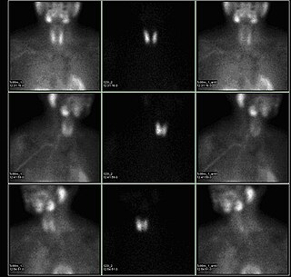
A sestamibi parathyroid scan is a procedure in nuclear medicine which is performed to localize parathyroid adenoma, which causes Hyperparathyroidism. Adequate localization of parathyroid adenoma allows the surgeon to use a minimally invasive surgical approach.
| | This article related to pathology is a stub. You can help Wikipedia by expanding it. |