The foramen lacerum is a triangular hole in the base of skull, located between the sphenoid, the apex of the petrous temporal and the basilar part of the occipital.

The pterygopalatine ganglion is a parasympathetic ganglion found in the pterygopalatine fossa. It is largely innervated by the greater petrosal nerve ; and its axons project to the lacrimal glands and nasal mucosa. The flow of blood to the nasal mucosa, in particular the venous plexus of the conchae, is regulated by the pterygopalatine ganglion and heats or cools the air in the nose. It is one of four parasympathetic ganglia of the head and neck, the others being the submandibular ganglion, otic ganglion, and ciliary ganglion.

The cavernous sinus within the human head, is one of the dural venous sinuses creating a cavity called the lateral sellar compartment bordered by the temporal bone of the skull and the sphenoid bone, lateral to the sella turcica.

The superior ophthalmic vein begins at the inner angle of the orbit in a vein named the nasofrontal which communicates anteriorly with the angular vein; it pursues the same course as the ophthalmic artery, and receives tributaries corresponding to the branches of that vessel.
The inferior ophthalmic vein begins in a venous network at the forepart of the floor and medial wall of the orbit; it receives some vorticose veins and other veins from the inferior rectus muscle, inferior oblique muscle, lacrimal sac and eyelids, runs backward in the lower part of the orbit lying above the inferior rectus and divides into two branches.

The pterygoid plexus is a venous plexus of considerable size, and is situated between the temporalis muscle and lateral pterygoid muscle, and partly between the two pterygoid muscles.
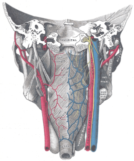
The pharyngeal veins begin in the pharyngeal plexus on the outer surface of the pharynx, and, after receiving some posterior meningeal veins and the vein of the pterygoid canal, end in the internal jugular.
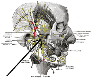
The medial pterygoid nerve is a branch of the mandibular nerve that innervates the medial pterygoid muscle, tensor veli palatini and tensor tympani.

The rectal venous plexus surrounds the rectum, and communicates in front with the vesical venous plexus in the male, and the vaginal venous plexus in the female.

The pharyngeal branch of the vagus nerve, the principal motor nerve of the pharynx, arises from the upper part of the ganglion nodosum, and consists principally of filaments from the cranial portion of the accessory nerve.
The anterior facial vein receives a branch of considerable size, the deep facial vein, from the pterygoid venous plexus.

The prostatic veins form a well-marked prostatic plexus which lies partly in the fascial sheath of the prostate and partly between the sheath and the prostatic capsule. It communicates with the pudendal and vesical plexuses.

The vaginal venous plexuses are placed at the sides of the vagina; they communicate with the uterine venous plexuses, vesical venous plexus, and rectal venous plexuses, and are drained by the vaginal veins, one on either side, into the hypogastric veins.
A venous plexus is a congregation of multiple veins.
The posterior scrotal veins are veins which drain into the vesical venous plexus.
The posterior labial veins are veins which drain to the vesical venous plexus.

The following outline is provided as an overview of and topical guide to human anatomy:

The Infratemporal space is a fascial space of the head and neck. It is a potential space in the side of the head, and is paired on either side. It is located posterior to the maxilla, between the lateral pterygoid plate of the sphenoid bone medially and by the base of skull superiorly. The term is derived from infra- meaning below and temporal which refers to the temporalis muscle.
The infraorbital vein arises on the face by the union of several tributaries. Accompanied by the infraorbital artery and the infraorbital nerve, it passes posteriorly through the infraorbital foramen, infraorbital canal, and infraorbital groove. It drains through the inferior orbital fissure into the pterygoid venous plexus. It receives tributaries from structures that lie close to the floor of the orbit and communicates with the inferior ophthalmic vein.
The public domain consists of all the creative works to which no exclusive intellectual property rights apply. Those rights may have expired, been forfeited, expressly waived, or may be inapplicable.

Gray's Anatomy is an English language textbook of human anatomy originally written by Henry Gray and illustrated by Henry Vandyke Carter. Earlier editions were called Anatomy: Descriptive and Surgical, Anatomy of the Human Body and Gray's Anatomy: Descriptive and Applied, but the book's name is commonly shortened to, and later editions are titled, Gray's Anatomy. The book is widely regarded as an extremely influential work on the subject, and has continued to be revised and republished from its initial publication in 1858 to the present day. The latest edition of the book, the 41st, was published in September 2015.