Related Research Articles

Microtubules are polymers of tubulin that form part of the cytoskeleton and provide structure and shape to eukaryotic cells. Microtubules can be as long as 50 micrometres, as wide as 23 to 27 nm and have an inner diameter between 11 and 15 nm. They are formed by the polymerization of a dimer of two globular proteins, alpha and beta tubulin into protofilaments that can then associate laterally to form a hollow tube, the microtubule. The most common form of a microtubule consists of 13 protofilaments in the tubular arrangement.

The evolution of flagella is of great interest to biologists because the three known varieties of flagella – each represent a sophisticated cellular structure that requires the interaction of many different systems.

The cytoskeleton is a complex, dynamic network of interlinking protein filaments present in the cytoplasm of all cells, including those of bacteria and archaea. In eukaryotes, it extends from the cell nucleus to the cell membrane and is composed of similar proteins in the various organisms. It is composed of three main components, microfilaments, intermediate filaments and microtubules, and these are all capable of rapid growth or disassembly dependent on the cell's requirements.

In cell biology, the spindle apparatus refers to the cytoskeletal structure of eukaryotic cells that forms during cell division to separate sister chromatids between daughter cells. It is referred to as the mitotic spindle during mitosis, a process that produces genetically identical daughter cells, or the meiotic spindle during meiosis, a process that produces gametes with half the number of chromosomes of the parent cell.

FtsZ is a protein encoded by the ftsZ gene that assembles into a ring at the future site of bacterial cell division. FtsZ is a prokaryotic homologue of the eukaryotic protein tubulin. The initials FtsZ mean "Filamenting temperature-sensitive mutant Z." The hypothesis was that cell division mutants of E. coli would grow as filaments due to the inability of the daughter cells to separate from one another. FtsZ is found in almost all bacteria, many archaea, all chloroplasts and some mitochondria, where it is essential for cell division. FtsZ assembles the cytoskeletal scaffold of the Z ring that, along with additional proteins, constricts to divide the cell in two.
Tubulin in molecular biology can refer either to the tubulin protein superfamily of globular proteins, or one of the member proteins of that superfamily. α- and β-tubulins polymerize into microtubules, a major component of the eukaryotic cytoskeleton. Microtubules function in many essential cellular processes, including mitosis. Tubulin-binding drugs kill cancerous cells by inhibiting microtubule dynamics, which are required for DNA segregation and therefore cell division.

RecA is a 38 kilodalton protein essential for the repair and maintenance of DNA. A RecA structural and functional homolog has been found in every species in which one has been seriously sought and serves as an archetype for this class of homologous DNA repair proteins. The homologous protein is called RAD51 in eukaryotes and RadA in archaea.
The Ames strain is one of 89 known strains of the anthrax bacterium. It was isolated from a diseased 14-month-old Beefmaster heifer that died in Sarita, Texas in 1981. The strain was isolated at the Texas Veterinary Medical Diagnostic Laboratory and a sample was sent to the United States Army Medical Research Institute of Infectious Diseases (USAMRIID). Researchers at USAMRIID mistakenly believed the strain came from Ames, Iowa because the return address on the package was the USDA's National Veterinary Services Laboratories in Ames and mislabeled the specimen.
The bacterium, despite its simplicity, contains a well-developed cell structure which is responsible for some of its unique biological structures and pathogenicity. Many structural features are unique to bacteria and are not found among archaea or eukaryotes. Because of the simplicity of bacteria relative to larger organisms and the ease with which they can be manipulated experimentally, the cell structure of bacteria has been well studied, revealing many biochemical principles that have been subsequently applied to other organisms.
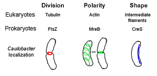
The prokaryotic cytoskeleton is the collective name for all structural filaments in prokaryotes. It was once thought that prokaryotic cells did not possess cytoskeletons, but advances in visualization technology and structure determination led to the discovery of filaments in these cells in the early 1990s. Not only have analogues for all major cytoskeletal proteins in eukaryotes been found in prokaryotes, cytoskeletal proteins with no known eukaryotic homologues have also been discovered. Cytoskeletal elements play essential roles in cell division, protection, shape determination, and polarity determination in various prokaryotes.
PcrA, standing for plasmid copy reduced is a helicase that was originally discovered in a screen for chromosomally encoded genes that are affected in plasmid rolling circle replication in the Gram-positive pathogen Staphylococcus aureus.

Bacillus anthracis is a gram-positive and rod-shaped bacterium that causes anthrax, a deadly disease to livestock and, occasionally, to humans. It is the only permanent (obligate) pathogen within the genus Bacillus. Its infection is a type of zoonosis, as it is transmitted from animals to humans. It was discovered by a German physician Robert Koch in 1876, and became the first bacterium to be experimentally shown as a pathogen. The discovery was also the first scientific evidence for the germ theory of diseases.

The AB toxins are two-component protein complexes secreted by a number of pathogenic bacteria, though there is a pore-forming AB toxin found the eggs of a snail. They can be classified as Type III toxins because they interfere with internal cell function. They are named AB toxins due to their components: the "A" component is usually the "active" portion, and the "B" component is usually the "binding" portion. The "A" subunit possesses enzyme activity, and is transferred to the host cell following a conformational change in the membrane-bound transport "B" subunit. These proteins consist of two independent polypeptides, which correspond to the A/B subunit moieties. The enzyme component (A) enters the cell through endosomes produced by the oligomeric binding/translocation protein (B), and prevents actin polymerisation through ADP-ribosylation of monomeric G-actin.
Fission, in biology, is the division of a single entity into two or more parts and the regeneration of those parts to separate entities resembling the original. The object experiencing fission is usually a cell, but the term may also refer to how organisms, bodies, populations, or species split into discrete parts. The fission may be binary fission, in which a single organism produces two parts, or multiple fission, in which a single entity produces multiple parts.
The parDE type II toxin-antitoxin system is one example of the bacterial toxin-antitoxin (TA) systems that encode two proteins, one a potent inhibitor of cell proliferation (toxin) and the other its specific antidote (antitoxin). These systems preferentially guarantee growth of plasmid-carrying daughter cells in a bacterial population by killing newborn bacteria that have not inherited a plasmid copy at cell division.
The parABS system is a broadly conserved molecular mechanism for plasmid partitioning and chromosome segregation in bacteria. Originally identified as a genetic element required for faithful partitioning of low-copy-number plasmids, it consists of three components: the ParA ATPase, the ParB DNA-binding protein, and the cis-acting parS sequence. The parA and parB genes are typically found in the same operon, with parS elements located within or adjacent to this operon. Collectively, these components function to ensure accurate partitioning of plasmids or whole chromosomes between bacterial daughter cells prior to cell division.
A plasmid partition system is a mechanism that ensures the stable inheritance of plasmids during bacterial cell division. Each plasmid has its independent replication system which controls the number of copies of the plasmid in a cell. The higher the copy number is, the more likely the two daughter cells will contain the plasmid. Generally, each molecule of plasmid diffuses randomly, so the probability of having a plasmid-less daughter cell is 21−N, where N is the number of copies. For instance, if there are 2 copies of a plasmid in a cell, there is a 50% chance of having one plasmid-less daughter cell. However, high-copy number plasmids have a cost for the hosting cell. This metabolic burden is lower for low-copy plasmids, but those have a higher probability of plasmid loss after a few generations. To control vertical transmission of plasmids, in addition to controlled-replication systems, bacterial plasmids use different maintenance strategies, such as multimer resolution systems, post-segregational killing systems, and partition systems.
The ParMRC system is a mechanism for sorting DNA plasmids to opposite ends of a bacterial cell during cell division. It has three components: ParM, an actin-like protein that forms a long filament to push two plasmids apart, ParR, which binds the plasmid to ParM and generates the ParM filament, and parC, which is a DNA sequence on the plasmid that anchors ParR to itself.

The divisome is a protein complex in bacteria that is responsible for cell division, constriction of inner and outer membranes during division, and peptidoglycan (PG) synthesis at the division site. The divisome is a membrane protein complex with proteins on both sides of the cytoplasmic membrane. In gram-negative cells it is located in the inner membrane. The divisome is nearly ubiquitous in bacteria although its composition may vary between species.
Prosthecobacter is a genus of bacteria from the phylum Verrucomicrobiota with a distinctive characteristic; the presence of tubulin-like genes. Tubulins, which are components of the microtubule, have never been observed in Gracilicutes before.
References
- Hayes, F; Barillà, D (Feb 2006). "The bacterial segrosome: a dynamic nucleoprotein machine for DNA trafficking and segregation". Nat Rev Microbiol. 4 (2): 133–43. doi:10.1038/nrmicro1342. PMID 16415929. S2CID 10082023.
- Hayes, F; Barillà, D (May 2006). "Assembling the bacterial segrosome". Trends Biochem Sci. 31 (5): 247–50. doi:10.1016/j.tibs.2006.03.002. PMID 16584885.
- Tinsley, E; Khan, SA (Apr 2006). "A novel FtsZ-like protein is involved in replication of the anthrax toxin-encoding pXO1 plasmid in Bacillus anthracis". J Bacteriol. 188 (8): 2829–35. doi:10.1128/jb.188.8.2829-2835.2006. PMC 1446996 . PMID 16585744.
- Larsen, RA; Cusumano, C; Fujioka, A; Lim-Fong, G; Patterson, P; Pogliano, J (Jun 2007). "Treadmilling of a prokaryotic tubulin-like protein, TubZ, required for plasmid stability in Bacillus thuringiensis". Genes Dev. 21 (11): 1340–52. doi:10.1101/gad.1546107. PMC 1877747 . PMID 17510284.
- Margolin, W (Aug 2007). "Bacterial cytoskeleton: not your run-of-the-mill tubulin". Curr. Biol. 17 (16): R633. doi:10.1016/j.cub.2007.06.020. PMC 4758210 . PMID 17714650.
- Anand, SP; Akhtar, P; Tinsley, E; Watkins, SC; Khan, SA (Feb 2008). "GTP-dependent polymerization of the tubulin-like RepX replication protein encoded by the pXO1 plasmid of Bacillus anthracis". Mol Microbiol. 67 (4): 881–90. doi: 10.1111/j.1365-2958.2007.06100.x . PMID 18179418.