Related Research Articles
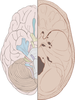
Cranial nerves are the nerves that emerge directly from the brain, of which there are conventionally considered twelve pairs. Cranial nerves relay information between the brain and parts of the body, primarily to and from regions of the head and neck, including the special senses of vision, taste, smell, and hearing.

A fontanelle is an anatomical feature of the infant human skull comprising any of the soft membranous gaps (sutures) between the cranial bones that make up the calvaria of a fetus or an infant. Fontanelles allow for rapid stretching and deformation of the neurocranium as the brain expands faster than the surrounding bone can grow. Premature complete ossification of the sutures is called craniosynostosis.

The ethmoid bone is an unpaired bone in the skull that separates the nasal cavity from the brain. It is located at the roof of the nose, between the two orbits. The cubical bone is lightweight due to a spongy construction. The ethmoid bone is one of the bones that make up the orbit of the eye.
The sphenoid bone is an unpaired bone of the neurocranium. It is situated in the middle of the skull towards the front, in front of the basilar part of the occipital bone. The sphenoid bone is one of the seven bones that articulate to form the orbit. Its shape somewhat resembles that of a butterfly or bat with its wings extended.
The occipital bone is a cranial dermal bone and the main bone of the occiput. It is trapezoidal in shape and curved on itself like a shallow dish. The occipital bone overlies the occipital lobes of the cerebrum. At the base of skull in the occipital bone, there is a large oval opening called the foramen magnum, which allows the passage of the spinal cord.

Choroid plexus cysts (CPCs) are cysts that occur within choroid plexus of the brain. They are the most common type of intraventricular cyst. The brain contains pockets or spaces called ventricles with a spongy layer of cells and blood vessels called the choroid plexus. This is in the middle of the fetal brain. The choroid plexus has the important function of producing cerebrospinal fluid. The fluid produced by the cells of the choroid plexus fills the ventricles and then flows around the brain and the spinal cord to provide a cushion of fluid around these structures.

Apert syndrome is a form of acrocephalosyndactyly, a congenital disorder characterized by malformations of the skull, face, hands and feet. It is classified as a branchial arch syndrome, affecting the first branchial arch, the precursor of the maxilla and mandible. Disturbances in the development of the branchial arches in fetal development create lasting and widespread effects.

Craniosynostosis is a condition in which one or more of the fibrous sutures in an infant skull prematurely fuses by turning into bone (ossification), thereby changing the growth pattern of the skull. Because the skull cannot expand perpendicular to the fused suture, it compensates by growing more in the direction parallel to the closed sutures. Sometimes the resulting growth pattern provides the necessary space for the growing brain, but results in an abnormal head shape and abnormal facial features. In cases in which the compensation does not effectively provide enough space for the growing brain, craniosynostosis results in increased intracranial pressure leading possibly to visual impairment, sleeping impairment, eating difficulties, or an impairment of mental development combined with a significant reduction in IQ.

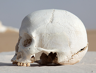
A skull fracture is a break in one or more of the eight bones that form the cranial portion of the skull, usually occurring as a result of blunt force trauma. If the force of the impact is excessive, the bone may fracture at or near the site of the impact and cause damage to the underlying structures within the skull such as the membranes, blood vessels, and brain.

A basilar skull fracture is a break of a bone in the base of the skull. Symptoms may include bruising behind the ears, bruising around the eyes, or blood behind the ear drum. A cerebrospinal fluid (CSF) leak occurs in about 20% of cases and can result in fluid leaking from the nose or ear. Meningitis is a complication in about 14% of cases. Other complications include cranial nerve or blood vessel injury.

The frontal suture is a fibrous joint that divides the two halves of the frontal bone of the skull in infants and children. Typically, it completely fuses between three and nine months of age, with the two halves of the frontal bone being fused together. It is also called the metopic suture, although this term may also refer specifically to a persistent frontal suture.

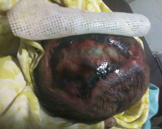
Encephalocele is a neural tube defect characterized by sac-like protrusions of the brain and the membranes that cover it through openings in the skull. These defects are caused by failure of the neural tube to close completely during fetal development. Encephaloceles cause a groove down the middle of the skull, or between the forehead and nose, or on the back side of the skull. The severity of encephalocele varies, depending on its location.

The foramen rotundum is a circular hole in the sphenoid bone that connects the middle cranial fossa and the pterygopalatine fossa.
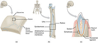
In anatomy, fibrous joints are joints connected by fibrous tissue, consisting mainly of collagen. These are fixed joints where bones are united by a layer of white fibrous tissue of varying thickness. In the skull the joints between the bones are called sutures. Such immovable joints are also referred to as synarthroses.

Artificial cranial deformation or modification, head flattening, or head binding is a form of body alteration in which the skull of a human being is deformed intentionally. It is done by distorting the normal growth of a child's skull by applying force. Flat shapes, elongated ones, rounded ones, and conical ones are among those chosen or valued in various cultures. Typically, the shape alteration is carried out on an infant, as the skull is most pliable at this time. In a typical case, headbinding begins approximately a month after birth and continues for about six months.

Obstetrical forceps are an instrument that can be used to assist in the delivery of a baby as an alternative to the ventouse method.
Acrania is a rare congenital disorder that occurs in the human fetus in which the flat bones in the cranial vault are either completely or partially absent. The cerebral hemispheres develop completely but abnormally. The condition is frequently, though not always, associated with anencephaly. The fetus is said to suffer from acrania if it meets the following criteria: the foetus should have a perfectly normal facial bone, a normal cervical column but without the fetal skull and a volume of brain tissue equivalent to at least one third of the normal brain size.

The endocranium in comparative anatomy is a part of the skull base in vertebrates and it represents the basal, inner part of the cranium. The term is also applied to the outer layer of the dura mater in human anatomy.
Cranial kinesis is the term for significant movement of skull bones relative to each other in addition to movement at the joint between the upper and lower jaw. It is usually taken to mean relative movement between the upper jaw and the braincase.
References
- ↑ THOMSON JL (February 1950). "The differential diagnosis of Spalding's sign". Br J Radiol. 23 (266): 122–4, illust. PMID 15409772.
- ↑ synd/2946 at Who Named It?
- ↑ A. B. Spalding. A pathogonomic sign of intra-uterine death. Surgery, Gynecology and Obstetrics, Chicago, 1922, 34: 754.
- ↑ 00509 at CHORUS
- ↑ The essentials of forensic medicine and toxicology. New delhi: Jaypee brothers. 2014. p. 438. ISBN 9789351525578.
- ↑ Olds' Maternal-Newborn Nursing, 8th edition, p. 1136
- ↑ S. A. Journal of Radiology, March 1964, OVERLAPPING OF THE FOETAL SKULL BONES IN BREECH PRESENTATION L. C. HANDLER, Department of Radiodiagnosis, Groote Schuur Hospital, Cape Town
| This human reproduction article is a stub. You can help Wikipedia by expanding it. |
| This medical sign article is a stub. You can help Wikipedia by expanding it. |