Surface-assisted laser desorption/ionization

Last updated

Surface-assisted laser desorption/ionization (SALDI) is a soft laser desorption technique used for mass spectrometry analysis of biomolecules, polymers, and small organic molecules. [1] [2] [3] [4] In its first embodiment Koichi Tanaka used a cobalt/glycerol liquid matrix [1] and subsequent applications included a graphite/glycerol liquid matrix as well as a solid surface of porous silicon. [3] The porous silicon represents the first matrix-free SALDI surface analysis allowing for facile detection of intact molecular ions, [3] [5] these porous silicon surfaces also facilitated the analysis of small molecules at the yoctomole level. [5] [6] At present laser desorption/ionization methods using other inorganic matrices such as nanomaterials are often regarded as SALDI variants. As an example, silicon nanowires [7] as well as Titania nanotube arrays (NTA) have been used as substrates to detect small molecules. [8] SALDI is used to detect proteins and protein-protein complexes. [9] A related method named "ambient SALDI" - which is a combination of conventional SALDI with ambient mass spectrometry incorporating the direct analysis real time (DART) ion source has also been demonstrated. [10] SALDI is considered one of the most important techniques in MS and has many applications. [11]

Contents

Schematic diagram of surface assisted laser desorption /ionization The blue circles represent the surface particles, the red circles represent the analyte molecules and the red circles with charges represent the charged analyte. SALDI.jpg
Schematic diagram of surface assisted laser desorption /ionization The blue circles represent the surface particles, the red circles represent the analyte molecules and the red circles with charges represent the charged analyte.

History

Koichi Tanaka performed the first successful LDI experiments on proteins. [1] Subsequently, Sunner and Chen used graphite particles of 2-150 μm in size as a substrate and solutions of analytes in glycerol to analyze low molecular weight analytes, peptides, and small proteins by surface-assisted laser desorption/ionization MS (SALDI-MS). [2] The technique was soon picked up by the Siuzdak lab who used nanostructured silicon surface for analyses. [3] Subsequent work on nanostructures included the addition of fluorinated "initiator" molecules onto the porous surface to enhance desorption/ionization, mass range, surface robustness, and sensitivity. [12] Other surface-based SALDI-MS approaches have also developed, including in 2000 where a thin layer of activated carbon particles fixed on aluminum support [13] Since the original 1999 nanostructured silicon [3] experiments, the research has largely focused on introducing novel nanomaterials as substrates, to enhance the sensitivity, broaden the mass range and expand the categories of samples that can be analyzed using this technique. [14]

SALDI was introduced as a promising method with potential applications in systems biology, particularly metabolomics. The introduction of nanomaterials as SALDI substrates attracted researchers in analytical chemistry. Such materials include carbon nanotubes (CNTs), metallic nanoparticles like Ag, Pt, Au, and nanostructured surfaces. This development of substrates allowed for further development of SALDI.

The development of desorption/ionization on silicon (DIOS)-MS [3] in particular, and subsequently nanostructure-initiator mass spectrometry (NIMS) [12] and nano-assisted laser desorption/ionization (NALDI), has also attracted the attention of analytical scientists. These methods have since become a benchmark for semiconductor-based SALDI research. [11] [14]

Basic principles

The main principle of SALDI relies on a medium that absorbs energy from a laser and then transfers the energy to the target sample. This class of techniques where the bulk of energy goes to the substrate instead of the sample molecules is known as soft ionization techniques. The development of SALDI started as a modification of matrix-assisted laser desorption/ionization (MALDI). The former technique suffered from ionization interference from the matrix molecules of MALDI. SALDI substituted an active surface of specific substrates, usually made of inorganic components, for the organic matrix of MALDI. [14]

SALDI is a three-stage process. The first stage is mainly concerned with mixing the samples with the substrate. In the second stage, the laser pulses are applied to the mix where the substrate absorbs the laser energy and transfers it to the sample molecules. In the final stage desorption and ionization occur and the potential difference accelerates produced ions into the mass analyzer.

Substrates

The substrate surface is playing a big role in adsorption, desorption, and ionization of the analyte molecules. This role is affected mainly by the chemical and physical properties of the substrate. Among these physical properties are the optical absorption coefficient, heat capacity and heat conductivity. [15]

1) The optical absorption coefficient: as this increases the ability of the substrate to absorb and generate more heat when absorb energy increases.

2) The heat capacity: as this decreases, the same amount of heat induces a larger temperature increase.

3) The heat conductivity: as this decreases, the substrate is better able to maintain the high temperature; therefore, the adsorption, desorption and ionization of the analytes occur more rapidly and effectively.

There are three classes of nanomaterials that are utilized in SALDI-MS. Namely, the carbon-based, semiconductor-based and metallic-based.

The forms of Carbon Nanotubes: Multiple or Single Wall Carbon Nanotube Nanotubos tipos.png
The forms of Carbon Nanotubes: Multiple or Single Wall Carbon Nanotube

Carbon nanotubes and carbon-based SALDI

The term carbon nanotube refers to a cylinder with a rolled graphene sheet. CNT can be single walled (SWNT) or multi-walled (MWNT). The SWNTs are perfect simulators of an ideal blackbody in the electromagnetic radiation ranging from the UV to far infrared. They exhibit better performance than former materials like super black, (a chemically etched nickel-phosphorus alloy). [16] This makes the CNT's a desired material for laser mass spectrometry applications. [17] That's why they attracted the researchers since discovery in the year 1991. [18]

Graphene as a surface material

Graphene is a type of popular carbon nanomaterial discovered in 2004. It has a large surface area that could effectively attach the analyte molecules. On the other hand, the efficiency of desorption/ionization for analytes on a layer of graphene can be enhanced by its simple monolayer structure and unique electronic properties. Polar compounds including amino acids, polyamines, anticancer drugs, and nucleosides can be successfully analyzed. In addition, nonpolar molecules can be analyzed with high resolution and sensitivity due to the hydrophobic nature of graphene itself. Compared with a conventional matrix, graphene exhibits a high desorption/ionization efficiency for nonpolar compounds. The graphene substrate functions as a substrate to trap analytes and it transfers energy to the analytes upon laser irradiation, which allows for the analytes to be readily desorbed/ionized and the interference of matrix to be eliminated. It has been demonstrated that the use of graphene as a substrate material avoids the fragmentation of analytes and provides good reproducibility and a high salt tolerance. [14]

Nanostructured semiconductor-based SALDI

Porous silicon as a substrate material

Porous silicon acted as an effective substrate for SALDI, its porous structure helped in trapping the analytes and its unique optical activity transferred effectively the laser energy to the adsorbate. [3] It was effective for analyzing wide range of biological small molecules. recently, a new technique named nanostructure Imaging mass spectrometry (NIMS) was introduced as a result of using explosive vaporization for desorption. [11]

The mechanism for porous silicon surface as a SALDI substrate involves three steps:

1)Adsorption: the analyte is adsorbed by porous silicon through Hydrogen bond formation using the silanol groups.

2) Electronic excitation:laser pulse excite the silicon producing free electrons and positive charges in the surface layer.this increase the acidity of silanol groups which donate the proton easily to analytes.

3) Thermal Activation: analytes are activated thermally and dissociated from the surface. [14]

Instrumentation

Schematic illustration of SALDI instrument Schematic illustration of SALDI instrument.JPG
Schematic illustration of SALDI instrument

SALDI as an improvement of MALDI, used the very similar instrument to that of MALDI. It employs a laser source for pulsed laser generation which is responsible for excitation of the sample mixture, which consists of the analyte and substrate materials. On the other side of the instrument, the mass analyzer which separates the analytes according to their mass-to-charge ratio (m/z) and the detector are located. Analytes are accelerated to the analyzer by applying potential difference. [14]

Combination with GC

Analytes were introduced from gas chromatography (GC) instrument which was coupled to the SALDI-MS instead of analytes being adsorbed on a solid substrate and directly ionized by means of a pulsed laser. Coupling to GC increased the efficiency of ionization and sensitivity. This was introduced for the first time in 2009 by Sunner et al. [19]

Ambient SALDI

Recently, researchers were able to analyze in ambient conditions as a result of the involvement of DART ion source into the SALDI-MS system. [10]

Applications

Forensics

Forensic investigation owes DIOS the favor of producing evidence in contraceptive polymers in an alleged sexual assault case that could've never been made by any other analytical technique. [20] [21] Moreover, Pihlainen K. et al. reported that the technique showed great promise in the forensic analysis of illicit drugs. They also reported that the interference was diminished by using this technique. [22] [23]

In addition, another report stated that DIOS identified 11 impurities. [24] Profiling the impurities was expected to lead to their origin. Eight years later, the authors published another report and mentioned that the technique identified the catecholamines in a human peripheral blood lymphocyte extract. [25] Also the quantitation of salicylate in human serum was proved using the DIOS-MS in negative ion mode. [26]

Biomedical

Thomas et al. worked on a group of enzyme systems, were able to achieve monitoring and direct analysis of enzyme-catalyzed reaction by DIOS-MS. One famous result was the reaction of acetylcholineesterase (AChE) with acetylcholine producing choline. This approach gained more fame when showed the ability to detect the selectivity of different enzyme inhibitors. The study started with hyperzine A, tacrine, and 2,6-dimethoxyphenyl-N-butylcarbamate, which are all inhibitors of AChE. The Inhibitor constant (ki) value of each of the inhibitors was found to be an important factor of their inhibition potentials. DIOS-MS has another advantage over MALDI, it can detect additional information in the low-mass region of mass spectrum as it can detect peptide peaks, and also identify post-translation modifications. These capabilities have great applications in protein identification with more confidence. [11]

Clinical diagnosis

The DIOS-MS technique was employed as a novel technique for patient diagnosis by examining the patients' plasma. The study focused on patients with polycystic ovarian syndrome (POS) by comparing the DIOS metabolic profiles generated with those of healthy subjects. The information obtained can be used to estimate disease progression and the effect of medical treatment. [11]

Pharmaceutical

An interesting area for researchers is to mobilize or immobilize some target proteins. It's a requirement in drug development mechanisms as some proteins' binding partners are not discovered yet. This was achieved by Zou et al. by using the DIOS technique. They employed a Psi surface as a probe to immobilize a targeted protein.

Next, the test drugs with the trial binding partners were introduced and incubated the probe. The immobilized proteins were able to capture the drug molecules that had a high affinity for the targeted protein. The ones with low affinity were washed off. The next step was to identify the captured drug molecules, and this was done by the SALDI analysis. The process offers great selectivity in testing drug candidates; it filters out the weak candidates and picks the most effective ones. It's not limited to proteins, it can work with macro biological molecules like DNA and RNA. [11]

Another famous test was done with hemoglobin. In this test, a hemoglobin-modified surface was employed. The target was to identify the non-covalent binding between hemoglobin and relevant chemicals. Among 13 different chemicals that included antimicrobials, insecticides, fungicides and herbicides, only triphenyltin chloride succeeded in binding hemoglobin strongly. This was a practical warning of the high toxicity of this material relative to other tested compounds.

Biochemical

Metabolic profiling

With increasing work and research in metabolomics, new techniques were needed for introducing novel study approaches. SALDI-MS and the family of direct analysis mass spectrometry (DAMS) were introduced as novel approaches in metabolomics. Goodacre et al. employed the DIOS-MS technique to study the yeast. They portrayed the yeast metabolites secreted by yeast showing that the metabolic "footprinting" of yeast is achievable. In a prior research work, the same group employed direct infusion mass spectrometry (DIMS) with electrospray ionization. They studied the metabolic profiles of a large number of wild types and mutants; and mathematical techniques were employed for data analysis to determine potential biomarkers. [11]

Imaging mass spectrometry

SALDI has been employed for imaging of a mouse heart and brain tissues. [27] This was the first achieved SALDI-MS. As in SALDI, laser has to penetrate through the tissue and be absorbed by the layer underneath, thickness would be a limiting factor, and researchers were able to overcome this factor by introducing an organic matrix onto the tissue section. This was named matrix enhanced surface-assisted laser desorption/ionization mass spectrometry (ME-SALDI-MS) to account for the different processes employed in the technique and refer to the modification that enhanced the technique.

More work was done for imaging of drug molecule distribution in brain tissue, then the cholesterol distribution in brain tissue and the sucrose distribution in Gerbera jamesonii flower stem. Also biofluids for direct analysis of drug molecules and their metabolites has been investigated. [11]

See also

Fast atom bombardment

Related Research Articles

<span class="mw-page-title-main">Mass spectrometry</span> Analytical technique based on determining mass to charge ratio of ions

Mass spectrometry (MS) is an analytical technique that is used to measure the mass-to-charge ratio of ions. The results are presented as a mass spectrum, a plot of intensity as a function of the mass-to-charge ratio. Mass spectrometry is used in many different fields and is applied to pure samples as well as complex mixtures.

<span class="mw-page-title-main">Ion source</span> Device that creates charged atoms and molecules (ions)

An ion source is a device that creates atomic and molecular ions. Ion sources are used to form ions for mass spectrometers, optical emission spectrometers, particle accelerators, ion implanters and ion engines.

<span class="mw-page-title-main">Koichi Tanaka</span> Japanese electrical engineer (born 1959)

Koichi Tanaka is a Japanese electrical engineer who shared the Nobel Prize in Chemistry in 2002 for developing a novel method for mass spectrometric analyses of biological macromolecules with John Bennett Fenn and Kurt Wüthrich.

<span class="mw-page-title-main">Matrix-assisted laser desorption/ionization</span> Ionization technique

In mass spectrometry, matrix-assisted laser desorption/ionization (MALDI) is an ionization technique that uses a laser energy-absorbing matrix to create ions from large molecules with minimal fragmentation. It has been applied to the analysis of biomolecules and various organic molecules, which tend to be fragile and fragment when ionized by more conventional ionization methods. It is similar in character to electrospray ionization (ESI) in that both techniques are relatively soft ways of obtaining ions of large molecules in the gas phase, though MALDI typically produces far fewer multi-charged ions.

Surface-enhanced laser desorption/ionization (SELDI) is a soft ionization method in mass spectrometry (MS) used for the analysis of protein mixtures. It is a variation of matrix-assisted laser desorption/ionization (MALDI). In MALDI, the sample is mixed with a matrix material and applied to a metal plate before irradiation by a laser, whereas in SELDI, proteins of interest in a sample become bound to a surface before MS analysis. The sample surface is a key component in the purification, desorption, and ionization of the sample. SELDI is typically used with time-of-flight (TOF) mass spectrometers and is used to detect proteins in tissue samples, blood, urine, or other clinical samples, however, SELDI technology can potentially be used in any application by simply modifying the sample surface.

Soft laser desorption (SLD) is laser desorption of large molecules that results in ionization without fragmentation. "Soft" in the context of ion formation means forming ions without breaking chemical bonds. "Hard" ionization is the formation of ions with the breaking of bonds and the formation of fragment ions.

<span class="mw-page-title-main">MALDI imaging</span>

MALDI mass spectrometry imaging (MALDI-MSI) is the use of matrix-assisted laser desorption ionization as a mass spectrometry imaging technique in which the sample, often a thin tissue section, is moved in two dimensions while the mass spectrum is recorded. Advantages, like measuring the distribution of a large amount of analytes at one time without destroying the sample, make it a useful method in tissue-based study.

<span class="mw-page-title-main">Protein mass spectrometry</span> Application of mass spectrometry

Protein mass spectrometry refers to the application of mass spectrometry to the study of proteins. Mass spectrometry is an important method for the accurate mass determination and characterization of proteins, and a variety of methods and instrumentations have been developed for its many uses. Its applications include the identification of proteins and their post-translational modifications, the elucidation of protein complexes, their subunits and functional interactions, as well as the global measurement of proteins in proteomics. It can also be used to localize proteins to the various organelles, and determine the interactions between different proteins as well as with membrane lipids.

<span class="mw-page-title-main">Desorption electrospray ionization</span>

Desorption electrospray ionization (DESI) is an ambient ionization technique that can be coupled to mass spectrometry (MS) for chemical analysis of samples at atmospheric conditions. Coupled ionization sources-MS systems are popular in chemical analysis because the individual capabilities of various sources combined with different MS systems allow for chemical determinations of samples. DESI employs a fast-moving charged solvent stream, at an angle relative to the sample surface, to extract analytes from the surfaces and propel the secondary ions toward the mass analyzer. This tandem technique can be used to analyze forensics analyses, pharmaceuticals, plant tissues, fruits, intact biological tissues, enzyme-substrate complexes, metabolites and polymers. Therefore, DESI-MS may be applied in a wide variety of sectors including food and drug administration, pharmaceuticals, environmental monitoring, and biotechnology.

Mass spectrometry imaging (MSI) is a technique used in mass spectrometry to visualize the spatial distribution of molecules, as biomarkers, metabolites, peptides or proteins by their molecular masses. After collecting a mass spectrum at one spot, the sample is moved to reach another region, and so on, until the entire sample is scanned. By choosing a peak in the resulting spectra that corresponds to the compound of interest, the MS data is used to map its distribution across the sample. This results in pictures of the spatially resolved distribution of a compound pixel by pixel. Each data set contains a veritable gallery of pictures because any peak in each spectrum can be spatially mapped. Despite the fact that MSI has been generally considered a qualitative method, the signal generated by this technique is proportional to the relative abundance of the analyte. Therefore, quantification is possible, when its challenges are overcome. Although widely used traditional methodologies like radiochemistry and immunohistochemistry achieve the same goal as MSI, they are limited in their abilities to analyze multiple samples at once, and can prove to be lacking if researchers do not have prior knowledge of the samples being studied. Most common ionization technologies in the field of MSI are DESI imaging, MALDI imaging, secondary ion mass spectrometry imaging and Nanoscale SIMS (NanoSIMS).

Sample preparation for mass spectrometry is used for the optimization of a sample for analysis in a mass spectrometer (MS). Each ionization method has certain factors that must be considered for that method to be successful, such as volume, concentration, sample phase, and composition of the analyte solution. Quite possibly the most important consideration in sample preparation is knowing what phase the sample must be in for analysis to be successful. In some cases the analyte itself must be purified before entering the ion source. In other situations, the matrix, or everything in the solution surrounding the analyte, is the most important factor to consider and adjust. Often, sample preparation itself for mass spectrometry can be avoided by coupling mass spectrometry to a chromatography method, or some other form of separation before entering the mass spectrometer. In some cases, the analyte itself must be adjusted so that analysis is possible, such as in protein mass spectrometry, where usually the protein of interest is cleaved into peptides before analysis, either by in-gel digestion or by proteolysis in solution.

<span class="mw-page-title-main">Laser spray ionization</span>

Laser spray ionization refers to one of several methods for creating ions using a laser interacting with a spray of neutral particles or ablating material to create a plume of charged particles. The ions thus formed can be separated by m/z with mass spectrometry. Laser spray is one of several ion sources that can be coupled with liquid chromatography-mass spectrometry for the detection of larger molecules.

<span class="mw-page-title-main">Matrix-assisted laser desorption electrospray ionization</span>

Matrix-assisted laser desorption electrospray ionization (MALDESI) was first introduced in 2006 as a novel ambient ionization technique which combines the benefits of electrospray ionization (ESI) and matrix-assisted laser desorption/ionization (MALDI). An infrared (IR) or ultraviolet (UV) laser can be utilized in MALDESI to resonantly excite an endogenous or exogenous matrix. The term 'matrix' refers to any molecule that is present in large excess and absorbs the energy of the laser, thus facilitating desorption of analyte molecules. The original MALDESI design was implemented using common organic matrices, similar to those used in MALDI, along with a UV laser. The current MALDESI source employs endogenous water or a thin layer of exogenously deposited ice as the energy-absorbing matrix where O-H symmetric and asymmetric stretching bonds are resonantly excited by a mid-IR laser.

<span class="mw-page-title-main">Desorption atmospheric pressure photoionization</span>

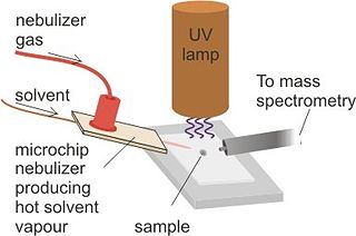
Desorption atmospheric pressure photoionization (DAPPI) is an ambient ionization technique for mass spectrometry that uses hot solvent vapor for desorption in conjunction with photoionization. Ambient Ionization techniques allow for direct analysis of samples without pretreatment. The direct analysis technique, such as DAPPI, eliminates the extraction steps seen in most nontraditional samples. DAPPI can be used to analyze bulkier samples, such as, tablets, powders, resins, plants, and tissues. The first step of this technique utilizes a jet of hot solvent vapor. The hot jet thermally desorbs the sample from a surface. The vaporized sample is then ionized by the vacuum ultraviolet light and consequently sampled into a mass spectrometer. DAPPI can detect a range of both polar and non-polar compounds, but is most sensitive when analyzing neutral or non-polar compounds. This technique also offers a selective and soft ionization for highly conjugated compounds.

<span class="mw-page-title-main">Capillary electrophoresis–mass spectrometry</span>

Capillary electrophoresis–mass spectrometry (CE–MS) is an analytical chemistry technique formed by the combination of the liquid separation process of capillary electrophoresis with mass spectrometry. CE–MS combines advantages of both CE and MS to provide high separation efficiency and molecular mass information in a single analysis. It has high resolving power and sensitivity, requires minimal volume and can analyze at high speed. Ions are typically formed by electrospray ionization, but they can also be formed by matrix-assisted laser desorption/ionization or other ionization techniques. It has applications in basic research in proteomics and quantitative analysis of biomolecules as well as in clinical medicine. Since its introduction in 1987, new developments and applications have made CE-MS a powerful separation and identification technique. Use of CE–MS has increased for protein and peptides analysis and other biomolecules. However, the development of online CE–MS is not without challenges. Understanding of CE, the interface setup, ionization technique and mass detection system is important to tackle problems while coupling capillary electrophoresis to mass spectrometry.

<span class="mw-page-title-main">Ambient ionization</span>

Ambient ionization is a form of ionization in which ions are formed in an ion source outside the mass spectrometer without sample preparation or separation. Ions can be formed by extraction into charged electrospray droplets, thermally desorbed and ionized by chemical ionization, or laser desorbed or ablated and post-ionized before they enter the mass spectrometer.

<span class="mw-page-title-main">Desorption/ionization on silicon</span> Soft laser desorption method

Desorption/ionization on silicon (DIOS) is a soft laser desorption method used to generate gas-phase ions for mass spectrometry analysis. DIOS is considered the first surface-based surface-assisted laser desorption/ionization (SALDI-MS) approach. Prior approaches were accomplished using nanoparticles in a matrix of glycerol, while DIOS is a matrix-free technique in which a sample is deposited on a nanostructured surface and the sample desorbed directly from the nanostructured surface through the adsorption of laser light energy. DIOS has been used to analyze organic molecules, metabolites, biomolecules and peptides, and, ultimately, to image tissues and cells.

<span class="mw-page-title-main">Matrix-assisted ionization</span>

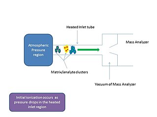
In mass spectrometry, matrix-assisted ionization is a low fragmentation (soft) ionization technique which involves the transfer of particles of the analyte and matrix sample from atmospheric pressure (AP) to the heated inlet tube connecting the AP region to the vacuum of the mass analyzer.

In mass spectrometry, a matrix is a compound that promotes the formation of ions. Matrix compounds are used in matrix-assisted laser desorption/ionization (MALDI), matrix-assisted ionization (MAI), and fast atom bombardment (FAB).

References

  1. 1 2 3 Tanaka, Koichi; Waki, Hiroaki; Ido, Yutaka; Akita, Satoshi; Yoshida, Yoshikazu; Yoshida, Tamio; Matsuo, T. (August 1988). "Protein and polymer analyses up tom/z 100 000 by laser ionization time-of-flight mass spectrometry". Rapid Communications in Mass Spectrometry. 2 (8): 151–153. Bibcode:1988RCMS....2..151T. doi:10.1002/rcm.1290020802. ISSN   0951-4198.
  2. 1 2 Sunner, Jan.; Dratz, Edward.; Chen, Yu-Chie. (1995). "Graphite surface-assisted laser desorption/ionization time-of-flight mass spectrometry of peptides and proteins from liquid solutions". Analytical Chemistry. 67 (23): 4335–4342. doi:10.1021/ac00119a021. PMID   8633776.
  3. 1 2 3 4 5 6 7 Wei, Jing; Buriak, Jillian M.; Siuzdak, Gary (May 1999). "Desorption–ionization mass spectrometry on porous silicon". Nature. 399 (6733): 243–246. Bibcode:1999Natur.399..243W. doi:10.1038/20400. ISSN   0028-0836. PMID   10353246. S2CID   4314372.
  4. Dattelbaum, Andrew M.; Iyer, Srinivas (2006-02-01). "Surface-assisted laser desorption/ionization mass spectrometry". Expert Review of Proteomics. 3 (1): 153–161. doi:10.1586/14789450.3.1.153. ISSN   1478-9450. PMID   16445359. S2CID   39538990.<!-https://zenodo.org/record/1235756-->
  5. 1 2 Trauger, Sunia A.; Go, Eden P.; Shen, Zhouxin; Apon, Junefredo V.; Compton, Bruce J.; Bouvier, Edouard S. P.; Finn, M. G.; Siuzdak, Gary (2004-08-01). "High Sensitivity and Analyte Capture with Desorption/Ionization Mass Spectrometry on Silylated Porous Silicon". Analytical Chemistry. 76 (15): 4484–4489. doi:10.1021/ac049657j. ISSN   0003-2700. PMID   15283591.
  6. Kurczy, Michael E.; Northen, Trent R.; Trauger, Sunia A.; Siuzdak, Gary (2015), He, Lin (ed.), "Nanostructure Imaging Mass Spectrometry: The Role of Fluorocarbons in Metabolite Analysis and Yoctomole Level Sensitivity", Mass Spectrometry Imaging of Small Molecules, Methods in Molecular Biology, vol. 1203, New York, NY: Springer, pp. 141–149, doi:10.1007/978-1-4939-1357-2_14, ISBN   978-1-4939-1357-2, PMC   4755109 , PMID   25361674
  7. Go, E. P.; Apon, J. V.; Luo, G.; Saghatelian, A.; Daniels, R. H.; Sahi, V.; Dubrow, R.; Cravatt, B. F.; Vertes, A. (March 2005). "Desorption/Ionization on Silicon Nanowires". Analytical Chemistry. 77 (6): 1641–1646. doi:10.1021/ac048460o. ISSN   0003-2700. PMID   15762567.
  8. Lo, Chun-Yuan; Lin, Jia-Yi; Chen, Wei-Yu; Chen, Cheng-Tai; Chen, Yu-Chie (2011-11-22). "Surface-assisted laser desorption/ionization mass spectrometry on titania nanotube arrays". Journal of the American Society for Mass Spectrometry. 19 (7): 1014–1020. doi: 10.1016/j.jasms.2008.04.025 . ISSN   1044-0305. PMID   18487059.
  9. Chen, Wen-Tsen; Chiang, Cheng-Kang; Lee, Chia-Hsin; Chang, Huan-Tsung (2012-02-06). "Using Surface-Assisted Laser Desorption/Ionization Mass Spectrometry to Detect Proteins and Protein–Protein Complexes". Analytical Chemistry. 84 (4): 1924–1930. doi:10.1021/ac202883q. PMID   22264081.
  10. 1 2 Zhang, Jialing; Li, Ze; Zhang, Chengsen; Feng, Baosheng; Zhou, Zhigui; Bai, Yu; Liu, Huwei (2012-03-13). "Graphite-Coated Paper as Substrate for High Sensitivity Analysis in Ambient Surface-Assisted Laser Desorption/Ionization Mass Spectrometry". Analytical Chemistry. 84 (7): 3296–3301. doi:10.1021/ac300002g. PMID   22380704.
  11. 1 2 3 4 5 6 7 8 Law, K. P.; Larkin, James R. (2010-08-21). "Recent advances in SALDI-MS techniques and their chemical and bioanalytical applications". Analytical and Bioanalytical Chemistry. 399 (8): 2597–2622. doi:10.1007/s00216-010-4063-3. ISSN   1618-2642. PMID   20730529. S2CID   36623535.
  12. 1 2 Northen, Trent R.; Yanes, Oscar; Northen, Michael T.; Marrinucci, Dena; Uritboonthai, Winnie; Apon, Junefredo; Golledge, Stephen L.; Nordström, Anders; Siuzdak, Gary (October 2007). "Clathrate nanostructures for mass spectrometry". Nature. 449 (7165): 1033–1036. Bibcode:2007Natur.449.1033N. doi:10.1038/nature06195. ISSN   0028-0836. PMID   17960240. S2CID   4404703.
  13. Han, Mei; Sunner, Jan (2000-07-01). "An activated carbon substrate surface for laser desorption mass spectrometry". Journal of the American Society for Mass Spectrometry. 11 (7): 644–649. doi:10.1016/S1044-0305(00)00129-X. ISSN   1044-0305. PMID   10883820. S2CID   22161820.
  14. 1 2 3 4 5 6 "OpenStax CNX". cnx.org. Retrieved 2016-04-05.
  15. Barron, Andrew R. (May 5, 2015). Physical Methods in Chemistry and Nano Science. OpenStax CNX.
  16. Mizuno, Kohei; Ishii, Juntaro; Kishida, Hideo; Hayamizu, Yuhei; Yasuda, Satoshi; Futaba, Don N.; Yumura, Motoo; Hata, Kenji (2009-04-14). "A black body absorber from vertically aligned single-walled carbon nanotubes". Proceedings of the National Academy of Sciences. 106 (15): 6044–6047. Bibcode:2009PNAS..106.6044M. doi: 10.1073/pnas.0900155106 . ISSN   0027-8424. PMC   2669394 . PMID   19339498.
  17. "Matrix-free Laser Desorption/Ionization on Vertically Aligned Carbon Nanotube Arrays". Bulletin of the Korean Chemical Society. 27 (4): 581–583. 2006. doi: 10.5012/bkcs.2006.27.4.581 .
  18. Iijima, Sumio (1991-11-07). "Helical microtubules of graphitic carbon". Nature. 354 (6348): 56–58. Bibcode:1991Natur.354...56I. doi:10.1038/354056a0. S2CID   4302490.
  19. Alimpiev, S.; Grechnikov, A.; Sunner, J.; Borodkov, A.; Karavanskii, V.; Simanovsky, Ya.; Nikiforov, S. (2008-12-24). "Gas Chromatography/Surface-Assisted Laser Desorption Ionization Mass Spectrometry of Amphetamine-like Compounds". Analytical Chemistry. 81 (3): 1255–1261. doi:10.1021/ac802176j. PMID   19108633.
  20. Thomas, John J.; Shen, Zhouxin; Blackledge, Robert; Siuzdak, Gary (2001-09-05). "Desorption–ionization on silicon mass spectrometry: an application in forensics". Analytica Chimica Acta. 442 (2): 183–190. doi:10.1016/S0003-2670(01)01107-2.
  21. Shen, Zhouxin; Thomas, John J.; Siuzdak, Gary; Blackledge, Robert D. (2004). "A Case Study on Forensic Polymer Analysis by DIOS-MS: The Suspect Who Gave Us the SLIP®". Journal of Forensic Sciences. 49 (5): 1–8. doi:10.1520/jfs2004026.
  22. Pihlainen, Katja; Grigoras, Kestutis; Franssila, Sami; Ketola, Raimo; Kotiaho, Tapio; Kostiainen, Risto (2005-04-01). "Analysis of amphetamines and fentanyls by atmospheric pressure desorption/ionization on silicon mass spectrometry and matrix-assisted laser desorption/ionization mass spectrometry and its application to forensic analysis of drug seizures". Journal of Mass Spectrometry. 40 (4): 539–545. Bibcode:2005JMSp...40..539P. doi:10.1002/jms.831. ISSN   1096-9888. PMID   15712357.
  23. Guinan, Taryn; Kirkbride, Paul; Pigou, Paul E.; Ronci, Maurizio; Kobus, Hilton; Voelcker, Nicolas H. (2015-11-01). "Surface-assisted laser desorption ionization mass spectrometry techniques for application in forensics". Mass Spectrometry Reviews. 34 (6): 627–640. Bibcode:2015MSRv...34..627G. doi:10.1002/mas.21431. ISSN   1098-2787. PMID   24916100.
  24. Kraj, Agnieszka; Świst, Małgorzta; Strugala, Anna; Parczewski, Andrzej; Silberring, Jerzy (2006). "Fingerprinting of 3, 4-methylenedioxymethamphetamine (MDMA) markers by desorption/ionization on porous silicon". European Journal of Mass Spectrometry. 12 (1): 253–9. doi:10.1255/ejms.811. PMID   17057282. S2CID   39575559.
  25. Kraj, Agnieszka; Jarzebinska, Justyna; Gorecka-Drzazga, Anna; Dziuban, Jan; Silberring, Jerzy (2006-07-15). "Identification of catecholamines in the immune system by desorption/ionization on silicon". Rapid Communications in Mass Spectrometry. 20 (13): 1969–1972. Bibcode:2006RCMS...20.1969K. doi:10.1002/rcm.2546. ISSN   1097-0231. PMID   16755607.
  26. Okuno, Shoji; Wada, Yoshinao (2005-08-01). "Measurement of serum salicylate levels by solid-phase extraction and desorption/ionization on silicon mass spectrometry". Journal of Mass Spectrometry. 40 (8): 1000–1004. Bibcode:2005JMSp...40.1000O. doi:10.1002/jms.859. ISSN   1096-9888. PMID   16059881.
  27. Liu, Qiang; Xiao, Yongsheng; Pagan-Miranda, Coral; Chiu, Yu Matthew; He, Lin (2011-11-22). "Metabolite imaging using matrix-enhanced surface-assisted laser desorption/ionization mass spectrometry (ME-SALDI-MS)". Journal of the American Society for Mass Spectrometry. 20 (1): 80–88. doi: 10.1016/j.jasms.2008.09.011 . ISSN   1044-0305. PMID   18926722.