The pancreas is an organ of the digestive system and endocrine system of vertebrates. In humans, it is located in the abdomen behind the stomach.

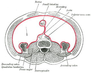
Edema, also spelled oedema or œdema, is an abnormal accumulation of fluid in the interstitium, located beneath the skin and in the cavities of the body, which can cause severe pain. Clinically, edema manifests as swelling. The amount of interstitial fluid is determined by the balance of fluid homeostasis and the increased secretion of fluid into the interstitium. The word is from Greek οἴδημα oídēma meaning "swelling". The condition is also known as dropsy.

Prune belly syndrome is a rare, genetic birth defect affecting about 1 in 40,000 births. About 97% of those affected are male. Prune belly syndrome is a congenital disorder of the urinary system, characterized by a triad of symptoms. The syndrome is named for the mass of wrinkled skin that is often present on the abdomen of those with the disorder.

The portal vein or hepatic portal vein is a blood vessel that carries blood from the gastrointestinal tract, gallbladder, pancreas and spleen to the liver. This blood contains nutrients and toxins extracted from digested contents. Approximately 75% of total liver blood flow is through the portal vein, with the remainder coming from the hepatic artery proper. The blood leaves the liver to the heart in the hepatic veins.

Cholecystitis is inflammation of the gallbladder. Symptoms include right upper abdominal pain, nausea, vomiting, and occasionally fever. Often gallbladder attacks precede acute cholecystitis. The pain lasts longer in cholecystitis than in a typical gallbladder attack. Without appropriate treatment, recurrent episodes of cholecystitis are common. Complications of acute cholecystitis include gallstone pancreatitis, common bile duct stones, or inflammation of the common bile duct.
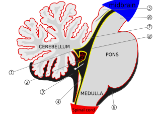
The choroid plexus is a plexus of cells that produces the cerebrospinal fluid in the ventricles of the brain. The choroid plexus consists of modified ependymal cells.
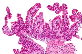
Whipple's disease is a rare, systemic infectious disease caused by the bacterium Tropheryma whipplei. First described by George Hoyt Whipple in 1907 and commonly considered a gastrointestinal disorder, Whipple's disease primarily causes malabsorption but may affect any part of the body including the heart, brain, joints, skin, lungs and the eyes. Weight loss, diarrhea, joint pain, and arthritis are common presenting symptoms, but the presentation can be highly variable and approximately 15% of patients do not have these classic signs and symptoms.
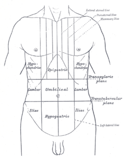
Abdominal pain, also known as a stomach ache, is a symptom associated with both non-serious and serious medical issues.
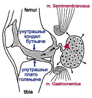
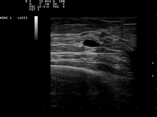
A Baker's cyst, also known as a popliteal cyst, is a type of fluid collection behind the knee. Often there are no symptoms. If symptoms do occur these may include swelling and pain behind the knee, or knee stiffness. If the cyst breaks open, pain may significantly increase with swelling of the calf. Rarely complications such as deep vein thrombosis, peripheral neuropathy, ischemia, or compartment syndrome may occur.

A chest radiograph, colloquially called a chest X-ray (CXR), or chest film, is a projection radiograph of the chest used to diagnose conditions affecting the chest, its contents, and nearby structures. Chest radiographs are the most common film taken in medicine.

Oral mucocele is a clinical term for two related phenomena: mucus extravasation phenomenon and mucus retention cyst. Other names include mucous extravasation cyst, mucous cyst of the oral mucosa, and mucous retention and extravasation phenomena.

Sialadenitis (sialoadenitis) is inflammation of salivary glands, usually the major ones, the most common being the parotid gland, followed by submandibular and sublingual glands. It should not be confused with sialadenosis (sialosis) which is a non-inflammatory enlargement of the major salivary glands.

Knee effusion occurs when excess synovial fluid accumulates in or around the knee joint. It has many common causes, including arthritis, injury to the ligaments or meniscus, or fluid collecting in the bursa, a condition known as prepatellar bursitis.

The periapical cyst is the most common odontogenic cyst. Periapical is defined as "the tissues surrounding the apex of the root of a tooth" and a cyst is "a pathological cavity lined by epithelium, having fluid or gaseous content that is not created by the accumulation of pus." Most frequently located in the maxillary anterior region, it is caused by pulpal necrosis secondary to dental caries or trauma. The cyst has lining that is derived from the epithelial cell rests of Malassez which proliferate to form the cyst. Highly common in the oral cavity, the periapical cyst is asymptomatic, but highly significant because a secondary infection can cause pain and damage. In radiographs, it appears a radiolucency around the apex of a tooth's root.

Calcifying odotogenic cyst is a benign odontogenic tumor of cystic type most likely to affect the anterior areas of the jaws. It is most common in people in their second to third decades but can be seen at almost any age. On radiographs, the calcifying odontogenic cyst appears as a unilocular radiolucency. In one-third of cases, an impacted tooth is involved. Microscopically, there are many cells that are described as "ghost cells", enlarged eosinophilic epithelial cells without nuclei.
A breast cyst is a fluid-filled sac within the breast. One breast can have one or more breast cysts. They are often described as round or oval lumps with distinct edges. In texture, a breast cyst usually feels like a soft grape or a water-filled balloon, but sometimes a breast cyst feels firm.

Choledochal cysts are congenital conditions involving cystic dilatation of bile ducts. They are uncommon in western countries but not as rare in East Asian nations like Japan and China.

A central nervous system cyst is a type of cyst that presents and affects part of the central nervous system (CNS). They are usually benign and filled with either cerebrospinal fluid, blood, or tumor cells. CNS cysts are classified into two categories: cysts that originate from non-central nervous system tissue, migrate to, and form on a portion of the CNS, and cysts that originate within central nervous system tissue itself. Within these two categories, there are many types of CNS cysts that have been identified from previous studies.
Peritoneal carcinomatosis (PC) is intraperitoneal dissemination (carcinosis) of any form of cancer that does not originate from the peritoneum itself. PC is most commonly seen in abdominopelvic malignancies. Computed tomography (CT) is particularly important for detailed preoperative assessment and evaluation of the radiological Peritoneal Cancer Index (PCI). The imaging findings vary from simple ascites to multifocal discrete nodules and infiltrative peritoneal masses. Various tumours and tumour like conditions can mimic PC. A systematic analysis of CT imaging features is helpful to narrow down the differential diagnosis, staging and effectively guiding the patient management.

Vaginal cysts are uncommon benign cysts that develop in the vaginal wall. The type of epithelial tissue lining a cyst is used to classify these growths. They can be congenital. They can present in childhood and adulthood. The most common type is the squamous inclusion cyst. It develops within vaginal tissue present at the site of an episiotomy or other vaginal surgical sites. In most instances they do not cause symptoms and present with few or no complications. A vaginal cyst can develop on the surface of the vaginal epithelium or in deeper layers. Often, they are found by the women herself and as an incidental finding during a routine pelvic examination. Vaginal cysts can mimic other structures that protrude from the vagina such as a rectocele and cystocele. Some cysts can be distinguished visually but most will need a biopsy to determine the type. Vaginal cysts can vary in size and can grow as large as 7 cm. Other cysts can be present on the vaginal wall though mostly these can be differentiated. Vaginal cysts can often be palpated (felt) by a clinician. Vaginal cysts are one type of vaginal mass, others include cancers and tumors. The prevalence of vaginal cysts is uncertain since many go unreported but it is estimated that 1 out of 200 women have a vaginal cyst. Vaginal cysts may initially be discovered during pregnancy and childbirth. These are then treated to provide an unobstructed delivery of the infant. Growths that originate from the urethra and other tissue can present as cysts of the vagina.