The face is the front of an animal's head that features three of the head's sense organs, the eyes, nose, and mouth, and through which animals express many of their emotions. The face is crucial for human identity, and damage such as scarring or developmental deformities affects the psyche adversely.

Standard anatomical terms of location deal unambiguously with the anatomy of animals, including humans.

The skull is a bony structure that forms the head in vertebrates. It supports the structures of the face and provides a protective cavity for the brain. The skull is composed of two parts: the cranium and the mandible. In the human, these two parts are the neurocranium and the viscerocranium or facial skeleton that includes the mandible as its largest bone. The skull forms the anterior most portion of the skeleton and is a product of cephalisation—housing the brain, and several sensory structures such as the eyes, ears, nose, and mouth. In humans these sensory structures are part of the facial skeleton.

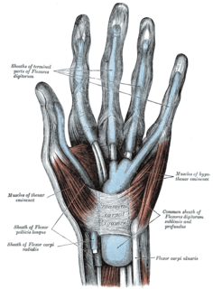
The thenar eminence refers to the group of muscles on the palm of the human hand at the base of the thumb. The skin overlying this region is the area stimulated when trying to elicit a palmomental reflex. The word thenar comes from Greek θέναρ (thenar), meaning 'palm of the hand'.

The subcutaneous tissue, also called the hypodermis, hypoderm, subcutis, or superficial fascia, is the lowermost layer of the integumentary system in vertebrates. The types of cells found in the hypodermis are fibroblasts, adipose cells, and macrophages. The hypodermis is derived from the mesoderm, but unlike the dermis, it is not derived from the dermatome region of the mesoderm. In arthropods, the hypodermis is an epidermal layer of cells that secretes the chitinous cuticle. The term also refers to a layer of cells lying immediately below the epidermis of plants.

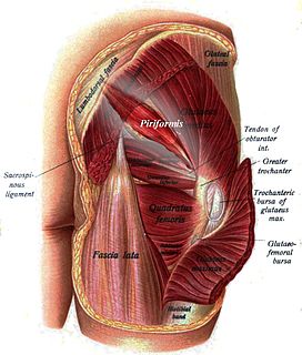
The piriformis is a muscle in the gluteal region of the lower limbs. It is one of the six muscles in the lateral rotator group.

The decapod crustacean, such as a crab, lobster, shrimp or prawn, is made up of 20 body segments grouped into two main body parts, the cephalothorax and the pleon (abdomen). Each segment may possess one pair of appendages, although in various groups these may be reduced or missing. They are, from head to tail:

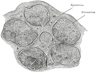
The epineurium is the outermost layer of dense irregular connective tissue surrounding a peripheral nerve. It usually surrounds multiple nerve fascicles as well as blood vessels which supply the nerve. Smaller branches of these blood vessels penetrate into the perineurium. In addition to blood vessels which supply the nerve, lymphocytes and fibroblasts are also present and contribute to the production of collagen fibers that form the backbone of the epineurium. In addition to providing structural support, lymphocytes and fibroblasts also play a vital role in maintenance and repair of the surrounding tissues.

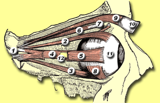
The levator palpebrae superioris is the muscle in the orbit that elevates the superior (upper) eyelid.

The iliopsoas refers to the joined psoas and the iliacus muscles. The two muscles are separate in the abdomen, but usually merge in the thigh. As such, they are usually given the common name iliopsoas. The iliopsoas muscle joins to the femur at the lesser trochanter, and acts as the strongest flexor of the hip.

The appendix of the epididymis is a small stalked appendage on the head of the epididymis. It is usually regarded as a detached efferent duct.

A cusp is a pointed, projecting, or elevated feature. In animals, it is usually used to refer to raised points on the crowns of teeth.

The premaxilla is one of a pair of small cranial bones at the very tip of the upper jaw of many animals, usually, but not always, bearing teeth. In humans, they are fused with the maxilla and usually termed as the incisive bone. Other terms used for this structure include premaxillary bone or os premaxillare, and intermaxillary bone or os intermaxillare.

The endocranium in comparative anatomy is a part of the skull base in vertebrates and it represents the basal, inner part of the cranium. The term is also applied to the outer layer of the dura mater in human anatomy.

A leg is a weight-bearing and locomotive anatomical structure, usually having a columnar shape. During locomotion, legs function as "extensible struts". The combination of movements at all joints can be modeled as a single, linear element capable of changing length and rotating about an omnidirectional "hip" joint.

Ape hand deformity, also known as simian hand, is a deformity in humans who cannot move the thumb away from the rest of the hand. It is an inability to abduct the thumb. Abduction of the thumb refers to the specific capacity to orient the thumb perpendicularly to the ventral (palmar) surface of the hand. Opposition refers specifically the ability to "swing" the first metacarpal such that the tip of the thumb may touch the distal end of the 5th phalanx and if we put the hand on the table as the palm upward the thumb can not point to the sky. The Ape Hand Deformity is caused by damage to the distal median nerve, and subsequent loss of opponens pollicis muscle function. The name "ape hand deformity" is misleading, as apes have opposable thumbs.
The dermatocranium is the portion of the cranium that is composed of dermal bone, as opposed to the endocranium and splanchnocranium, which are composed of endochondral bone. The dermatocranium comprises the skull roof, the facial skeleton, and—in fishes—the opercular bones.

Muscles are described using unique anatomical terminology according to their actions and structure.

In the vertebrate spinal column, each vertebra is an irregular bone with a complex structure composed of bone and some hyaline cartilage, the proportions of which vary according to the segment of the backbone and the species of vertebrate.


