Vitelline circulation

Last updated
Human embryo of 2.6 mm. Gray22.png
Human embryo of 2.6 mm.
Diagram of the vascular channels in a human embryo of the second week. (After Eternod.) The red lines are the dorsal aortae continued into the umbilical arteries. The red dotted lines are the ventral aortae, and the blue dotted lines the vitelline veins. Gray458.png
Diagram of the vascular channels in a human embryo of the second week. (After Eternod.) The red lines are the dorsal aortæ continued into the umbilical arteries. The red dotted lines are the ventral aortæ, and the blue dotted lines the vitelline veins.

Vitelline circulation refers to the system of blood flowing from the embryo to the yolk sac and back again.

Blood specialized bodily fluid in animals

Blood is a body fluid in humans and other animals that delivers necessary substances such as nutrients and oxygen to the cells and transports metabolic waste products away from those same cells.

Embryo multicellular diploid eukaryote in its earliest stage of development

An embryo is an early stage of development of a multicellular diploid eukaryotic organism. In general, in organisms that reproduce sexually, an embryo develops from a zygote, the single cell resulting from the fertilization of the female egg cell by the male sperm cell. The zygote possesses half the DNA from each of its two parents. In plants, animals, and some protists, the zygote will begin to divide by mitosis to produce a multicellular organism. The result of this process is an embryo.

Yolk sac membranous sac attached to an embryo, formed by cells of the hypoblast adjacent to the embryonic disk

The yolk sac is a membranous sac attached to an embryo, formed by cells of the hypoblast adjacent to the embryonic disk. This is alternatively called the umbilical vesicle by the Terminologia Embryologica (TE), though yolk sac is far more widely used. In humans, the yolk sac is important in early embryonic blood supply, and much of it is incorporated into the primordial gut during the fourth week of development.

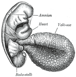
The yolk-sac is situated on the ventral aspect of the embryo; it is lined by endoderm, outside of which is a layer of mesoderm. It is filled with fluid, the vitelline fluid, which is utilized for the nourishment of the embryo during the earlier stages of its existence. Blood is conveyed to the wall of the sac by the vitelline arteries (a branch of the dorsal aorta), and after circulating through a wide-meshed capillary plexus, is returned by the vitelline veins to the tubular heart of the embryo. This constitutes the vitelline circulation, and by means of it nutritive material is absorbed from the yolk-sac and conveyed to the embryo.

Endoderm

Endoderm is one of the three primary germ layers in the very early embryo. The other two layers are the ectoderm and mesoderm, with the endoderm being the innermost layer. Cells migrating inward along the archenteron form the inner layer of the gastrula, which develops into the endoderm.

Mesoderm

In all bilaterian animals, the mesoderm is one of the three primary germ layers in the very early embryo. The other two layers are the ectoderm and endoderm, with the mesoderm as the middle layer between them.

Vitelline arteries

The vitelline arteries are the arterial counterpart to the vitelline veins. Like the veins, they play an important role in the vitelline circulation of blood to and from the yolk sac of a fetus. They are a branch of the dorsal aorta.

Related Research Articles

Circulatory system Organ system for circulating blood in animals

The circulatory system, also called the cardiovascular system or the vascular system, is an organ system that permits blood to circulate and transport nutrients, oxygen, carbon dioxide, hormones, and blood cells to and from the cells in the body to provide nourishment and help in fighting diseases, stabilize temperature and pH, and maintain homeostasis.

Amniotic sac

The amniotic sac, commonly called the bag of waters, sometimes the membranes, is the sac in which the fetus develops in amniotes. It is a thin but tough transparent pair of membranes that hold a developing embryo until shortly before birth. The inner of these fetal membranes, the amnion, encloses the amniotic cavity, containing the amniotic fluid and the fetus. The outer membrane, the chorion, contains the amnion and is part of the placenta. On the outer side, the amniotic sac is connected to the yolk sac, the allantois and, via the umbilical cord, to the placenta.

Amnion

The amnion is a membrane that closely covers the embryo when first formed. It fills with the amniotic fluid which causes the amnion to expand and become the amniotic sac which serves to provide a protective environment for the developing embryo or fetus. The amnion, along with the chorion, the yolk sac and the allantois form a protective sac around the embryo. In birds, reptiles and monotremes, the protective sac is enclosed in a shell. In marsupials and placental mammals, it is enclosed in a uterus.

Chorion

The chorion is the outermost fetal membrane around the embryo in mammals, birds and reptiles. It develops from an outer fold on the surface of the yolk sac, which lies outside the zona pellucida, known as the vitelline membrane in other animals. In insects it is developed by the follicle cells while the egg is in the ovary.

Allantois Embryonic structure

The allantois is a hollow sac-like structure filled with clear fluid that forms part of a developing amniote's conceptus. It helps the embryo exchange gases and handle liquid waste.

Vitelline duct

In the human embryo, the vitelline duct, also known as the vitellointestinal duct, the yolk stalk, the omphaloenteric duct, or the omphalomesenteric duct, is a long narrow tube that joins the yolk sac to the midgut lumen of the developing fetus. It appears at the end of the fourth week, when the yolk sac presents the appearance of a small pear-shaped vesicle.

Chorionic villi

Chorionic villi are villi that sprout from the chorion to provide maximal contact area with maternal blood.

Angioblast is the embryonic tissue from which blood vessels arise. Blood vessels first make their appearance in several scattered vascular areas that are developed simultaneously between the endoderm and the mesoderm of the yolk-sac, i. e., outside the body of the embryo. Here a new type of cell, the angioblast, is differentiated from the mesoderm.

Vitelline veins

The vitelline veins are veins that drain blood from the yolk sac.

Dorsal aorta

The dorsal aortae are paired embryological vessels which progress to form the descending aorta. The paired dorsal aortae arise from aortic arches that in turn arise from the aortic sac.

Anterior cardinal vein

The anterior cardinal veins contribute to the formation of the internal jugular veins and together with the common cardinal vein form the superior vena cava. In an anastomosis by anterior cardinal veins, the left brachiocephalic vein is produced.

Blood islands

Blood islands are structures around the developing embryo which lead to many different parts of the circulatory system. Blood islands arise external to the developing embryo on the umbilical vesicle, allantois, connecting stalk and chorion. They are also known as Pander's islands or Wolff's islands, after Heinz Christian Pander or Caspar Friedrich Wolff.

Lacrimal apparatus

The lacrimal apparatus is the physiological system containing the orbital structures for tear production and drainage.
It consists of:

Bilaminar blastocyst

Bilaminar blastocyst or Bilaminar disc refers to the epiblast and the hypoblast, evolved from the embryoblast. These two layers are sandwiched between two balloons: the primitive yolk sac and the amniotic cavity.

Human embryonic development process of cell division and cellular differentiation of the embryo that occurs during the early stages of development

Human embryonic development, or human embryogenesis, refers to the development and formation of the human embryo. It is characterised by the process of cell division and cellular differentiation of the embryo that occurs during the early stages of development. In biological terms, the development of the human body entails growth from a one-celled zygote to an adult human being. Fertilisation occurs when the sperm cell successfully enters and fuses with an egg cell (ovum). The genetic material of the sperm and egg then combine to form a single cell called a zygote and the germinal stage of development commences. Embryonic development in the human, covers the first eight weeks of development; at the beginning of the ninth week the embryo is termed a fetus. Human embryology is the study of this development during the first eight weeks after fertilisation. The normal period of gestation (pregnancy) is nine months or 38 weeks.

A choriovitelline placenta is a placenta formed by the yolk sac and chorion. In a choriovitelline placenta, the yolk sac fuses with the chorion and subsequently wrinkles develop that hold the embryo to the uterine wall and the embryo, thus forming the choriovitelline placenta. The chorionic blood vessels are connected with the vitelline blood vessel of the yolk sac.

References

This article incorporates text in the public domain from the 20th edition of Gray's Anatomy (1918)

The public domain consists of all the creative works to which no exclusive intellectual property rights apply. Those rights may have expired, been forfeited, expressly waived, or may be inapplicable.

<i>Grays Anatomy</i> English-language textbook of human anatomy

Gray's Anatomy is an English language textbook of human anatomy originally written by Henry Gray and illustrated by Henry Vandyke Carter. Earlier editions were called Anatomy: Descriptive and Surgical, Anatomy of the Human Body and Gray's Anatomy: Descriptive and Applied, but the book's name is commonly shortened to, and later editions are titled, Gray's Anatomy. The book is widely regarded as an extremely influential work on the subject, and has continued to be revised and republished from its initial publication in 1858 to the present day. The latest edition of the book, the 41st, was published in September 2015.