Related Research Articles

Fecal incontinence (FI), or in some forms, encopresis, is a lack of control over defecation, leading to involuntary loss of bowel contents — including flatus (gas), liquid stool elements and mucus, or solid feces. FI is a sign or a symptom, not a diagnosis. Incontinence can result from different causes and might occur with either constipation or diarrhea. Continence is maintained by several interrelated factors, including the anal sampling mechanism, and incontinence usually results from a deficiency of multiple mechanisms. The most common causes are thought to be immediate or delayed damage from childbirth, complications from prior anorectal surgery, altered bowel habits. An estimated 2.2% of community-dwelling adults are affected. However, reported prevalence figures vary. A prevalence of 8.39% among non-institutionalized U.S adults between 2005 and 2010 has been reported, and among institutionalized elders figures come close to 50%.

Anotia describes a rare congenital deformity that involves the complete absence of the auricle, the outer projected portion of the ear, and narrowing or absence of the ear canal. This contrasts with microtia, in which a small part of the auricle is present. Anotia and microtia may occur unilaterally or bilaterally. This deformity results in conductive hearing loss, deafness.

In the human brain, the diencephalon is a division of the forebrain. It is situated between the telencephalon and the midbrain. The diencephalon has also been known as the tweenbrain in older literature. It consists of structures that are on either side of the third ventricle, including the thalamus, the hypothalamus, the epithalamus and the subthalamus.

The male reproductive system consists of a number of sex organs that play a role in the process of human reproduction. These organs are located on the outside of the body, and within the pelvis.
The anal canal is the part that connects the rectum to the anus, located below the level of the pelvic diaphragm. It is located within the anal triangle of the perineum, between the right and left ischioanal fossa. As the final functional segment of the bowel, it functions to regulate release of excrement by two muscular sphincter complexes. The anus is the aperture at the terminal portion of the anal canal.

The internal anal sphincter, IAS, or sphincter ani internus is a ring of smooth muscle that surrounds about 2.5–4.0 cm of the anal canal. It is about 5 mm thick, and is formed by an aggregation of the smooth (involuntary) circular muscle fibers of the rectum. It terminates distally about 6 mm from the anal orifice.

Anal fistula is a chronic abnormal communication between the anal canal and the perianal skin. An anal fistula can be described as a narrow tunnel with its internal opening in the anal canal and its external opening in the skin near the anus. Anal fistulae commonly occur in people with a history of anal abscesses. They can form when anal abscesses do not heal properly.

The cloacal membrane is the membrane that covers the embryonic cloaca during the development of the urinary and reproductive organs.

The vaginal process is an embryonic developmental outpouching of the parietal peritoneum. It is present from around the 12th week of gestation, and commences as a peritoneal outpouching.

The pectinate line is a line which divides the upper two-thirds and lower third of the anal canal. Developmentally, this line represents the hindgut-proctodeum junction.

Anorectal abscess is an abscess adjacent to the anus. Most cases of perianal abscesses are sporadic, though there are certain situations which elevate the risk for developing the disease, such as diabetes mellitus, Crohn's disease, chronic corticosteroid treatment and others. It arises as a complication of paraproctitis. Ischiorectal, inter- and intrasphincteric abscesses have been described.

Human embryonic development or human embryogenesis is the development and formation of the human embryo. It is characterised by the processes of cell division and cellular differentiation of the embryo that occurs during the early stages of development. In biological terms, the development of the human body entails growth from a one-celled zygote to an adult human being. Fertilization occurs when the sperm cell successfully enters and fuses with an egg cell (ovum). The genetic material of the sperm and egg then combine to form the single cell zygote and the germinal stage of development commences. Human embryonic development covers the first eight weeks of development, which have 23 stages, called Carnegie stages. At the beginning of the ninth week, the embryo is termed a fetus. In comparison to the embryo, the fetus has more recognizable external features and a more complete set of developing organs.

A persistent cloaca is a symptom of a complex anorectal congenital disorder, in which the rectum, vagina, and urinary tract meet and fuse, creating a cloaca, a single common channel.
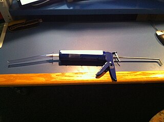
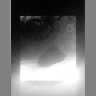
Defecography is a type of medical radiological imaging in which the mechanics of a patient's defecation are visualized in real time using a fluoroscope. The anatomy and function of the anorectum and pelvic floor can be dynamically studied at various stages during defecation.

In humans, the anus is the external opening of the rectum located inside the intergluteal cleft. Two sphincters control the exit of feces from the body during an act of defecation, which is the primary function of the anus. These are the internal anal sphincter and the external anal sphincter, which are circular muscles that normally maintain constriction of the orifice and which relax as required by normal physiological functioning. The inner sphincter is involuntary and the outer is voluntary. Above the anus is the perineum, which is also located beneath the vulva or scrotum.

Anorectal manometry (ARM) is a medical test used to measure pressures in the anus and rectum and to assess their function. The test is performed by inserting a catheter, that contains a probe embedded with pressure sensors, through the anus and into the rectum. Patients may be asked to perform certain maneuvers, such as coughing or attempting to defecate, to assess for pressure changes. Anorectal manometry is a safe and low risk procedure.
Anismus or dyssynergic defecation is the failure of normal relaxation of pelvic floor muscles during attempted defecation. It can occur in both children and adults, and in both men and women. It can be caused by physical defects or it can occur for other reasons or unknown reasons. Anismus that has a behavioral cause could be viewed as having similarities with parcopresis, or psychogenic fecal retention.
Endoanal ultrasound is a type of medical investigation which images the structures of the anal canal.
A keyhole defect is a term used in medicine and in the forensic sciences to refer to the shape of an anomalous feature or traumatic lesion caused by a gunshot wound.

Heart development, also known as cardiogenesis, refers to the prenatal development of the heart. This begins with the formation of two endocardial tubes which merge to form the tubular heart, also called the primitive heart tube. The heart is the first functional organ in vertebrate embryos.
References
- ↑ Sadler, Thomas W. (2011-12-15). Langman's Medical Embryology. Lippincott Williams & Wilkins. ISBN 9781451113426.
| | This developmental biology article is a stub. You can help Wikipedia by expanding it. |