The aorta is the main and largest artery in the human body, originating from the left ventricle of the heart and extending down to the abdomen, where it splits into two smaller arteries. The aorta distributes oxygenated blood to all parts of the body through the systemic circulation.

An artery is a blood vessel that takes blood away from the heart to all parts of the body. Most arteries carry oxygenated blood; the two exceptions are the pulmonary and the umbilical arteries, which carry deoxygenated blood to the organs that oxygenate it. The effective arterial blood volume is that extracellular fluid which fills the arterial system.
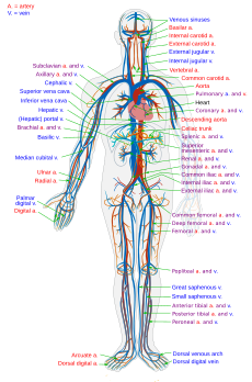
The blood vessels are a part of the circulatory system, and microcirculation, that transports blood throughout the human body. These vessels are designed to transport nutrients and oxygen to the tissues of the body. They also take waste and carbon dioxide and carry them away from the tissues and back to the heart. Blood vessels are needed to sustain life as all of the body’s tissues rely on their functionality.There are three major types of blood vessels: the arteries, which carry the blood away from the heart; the capillaries, which enable the actual exchange of water and chemicals between the blood and the tissues; and the veins, which carry blood from the capillaries back toward the heart. The word vascular, meaning relating to the blood vessels, is derived from the Latin vas, meaning vessel. Some structures -- such as cartilage, the epithelium, and the lens and cornea of the eye -- do not contain blood vessels and are labeled avascular.

Veins are blood vessels that carry blood toward the heart. Most veins carry deoxygenated blood from the tissues back to the heart; exceptions are the pulmonary and umbilical veins, both of which carry oxygenated blood to the heart. In contrast to veins, arteries carry blood away from the heart.

Atherosclerosis is a disease in which the inside of an artery narrows due to the build up of plaque. Initially, there are generally no symptoms. When severe, it can result in coronary artery disease, stroke, peripheral artery disease, or kidney problems, depending on which arteries are affected. Symptoms, if they occur, generally do not begin until middle age.
Macrovascular disease is a disease of any large (macro) blood vessels in the body. It is a disease of the large blood vessels, including the coronary arteries, the aorta, and the sizable arteries in the brain and in the limbs.

The coronary arteries are the arteries of the coronary circulation, which transports blood into and out of the cardiac muscle. They are mainly composed of the left and right coronary arteries, both of which give off branches. Coronary arteries can also be categorized as epicardial and microvascular.

A pseudoaneurysm, also known as a false aneurysm, is a collection of blood that forms between the two outer layers of an artery, the tunica media and the tunica adventitia. It is usually caused by a penetrating injury to the vessel, which then bleeds, but forms a space between the above two layers, rather than exiting the vessel. It may be pulsatile and can resemble a true aneurysm. A true aneurysm involves all three layers of the blood vessel. A dissecting aneurysm is when blood from the vessel lumen tracks between the two inner layers, the intima and the tunica media. This can cause blockage of the flow. A perivascular hematoma is a collection of blood that is external to the three vessel layers. Due to being close to the vessel, it can also be pulsatile, and can be mistaken for a pseudoaneurysm or aneurysm.

The vasa vasorum is a network of small blood vessels that supply the walls of large blood vessels, such as elastic arteries and large veins. The name derives from Latin, meaning 'the vessels of the vessels'.

Calcification is the accumulation of calcium salts in a body tissue. It normally occurs in the formation of bone, but calcium can be deposited abnormally in soft tissue, causing it to harden. Calcifications may be classified on whether there is mineral balance or not, and the location of the calcification. Calcification may also refer to the processes of normal mineral deposition in biological systems, such as the formation of stromatolites or mollusc shells.

Arteriolosclerosis is a form of cardiovascular disease involving hardening and loss of elasticity of arterioles or small arteries and is most often associated with hypertension and diabetes mellitus. Types include hyaline arteriolosclerosis and hyperplastic arteriolosclerosis, both involved with vessel wall thickening and luminal narrowing that may cause downstream ischemic injury. The following two terms whilst similar, are distinct in both spelling and meaning and may easily be confused with arteriolosclerosis.
A circulatory anastomosis is a connection between two blood vessels, such as between arteries, between veins or between an artery and a vein. Anastomoses between arteries and between veins result in a multitude of arteries and veins, respectively, serving the same volume of tissue. Such anastomoses occur normally in the body in the circulatory system, serving as backup routes for blood to flow if one link is blocked or otherwise compromised, but may also occur pathologically.

The tunica externa — also known as the tunica adventitia, or adventitia for short — is the outermost tunica (layer) of a blood vessel, surrounding the tunica media. It is mainly composed of collagen and, in arteries, is supported by external elastic lamina. The collagen serves to anchor the blood vessel to nearby organs, giving it stability.

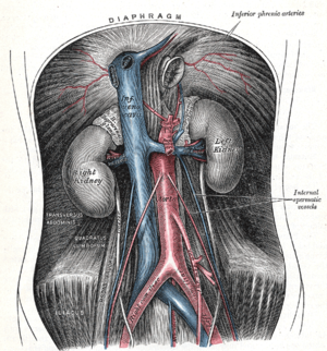
The scrotum is an anatomical male reproductive structure that consists of a suspended dual-chambered sack of skin and smooth muscle that is present in most terrestrial male mammals and located under the penis. One testis is typically lower than the other to avoid compression in the event of impact. The perineal raphe is a small, vertical, slightly raised ridge of scrotal skin under which is found the scrotal septum. It appears as a thin longitudinal line that runs front to back over the entire scrotum. The scrotum contains the external spermatic fascia, testes, epididymis, and ductus deferens. It is a distention of the perineum and carries some abdominal tissues into its cavity including the testicular artery, testicular vein, and pampiniform plexus. In humans and some other mammals, the scrotum becomes covered with pubic hair at puberty. The scrotum will usually tighten during penile erection and when exposed to cold temperature.

The internal elastic lamina or internal elastic lamella is a layer of elastic tissue that forms the outermost part of the tunica intima of blood vessels. It separates tunica intima from tunica media.
Penile ulltrasonography is medical ultrasonography of the penis. Ultrasound is an excellent method for the study of the penis, such as indicated in trauma, priapism, erectile dysfunction or suspected Peyronie's disease.