Articles related to anatomy include:

The lens is a transparent biconvex structure in the eye that, along with the cornea, helps to refract light to be focused on the retina. By changing shape, functions to change the focal length of the eye so that it can focus on objects at various distances, thus allowing a sharp real image of the object of interest to be formed on the retina. This adjustment of the lens is known as accommodation. Accommodation is similar to the focusing of a photographic camera via movement of its lenses. The lens is more flat on its anterior side than on its posterior side.
The vitreous body is the clear gel that fills the space between the lens and the retina of the eyeball of humans and other vertebrates. It is often referred to as the vitreous humor or simply "the vitreous".
A vulnerable plaque is a kind of atheromatous plaque – a collection of white blood cells and lipids in the wall of an artery – that is particularly unstable and prone to produce sudden major problems such as a heart attack or stroke.
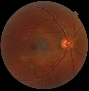
The optic disc or optic nerve head is the point of exit for ganglion cell axons leaving the eye. Because there are no rods or cones overlying the optic disc, it corresponds to a small blind spot in each eye.

The central retinal artery branches off the ophthalmic artery, running inferior to the optic nerve within its dural sheath to the eyeball.
The hyaloid artery is a branch of the ophthalmic artery, which is itself a branch of the internal carotid artery. It is contained within the optic stalk of the eye and extends from the optic disc through the vitreous humor to the lens. Usually fully regressed before birth, its purpose is to supply nutrients to the developing lens in the growing fetus.

The carotid sheath is an anatomical term for the fibrous connective tissue that surrounds the vascular compartment of the neck. It is part of the deep cervical fascia of the neck, below the superficial cervical fascia meaning the subcutaneous adipose tissue immediately beneath the skin.
ICD-10 is an international statistical classification used in health care and related industries.

The zonule of Zinn is a ring of fibrous strands forming a zonule that connects the ciliary body with the crystalline lens of the eye. These fibers are sometimes collectively referred to as the suspensory ligaments of the lens, as they act like suspensory ligaments.

A sphenoid wing meningioma is a benign brain tumor near the sphenoid bone.

The short posterior ciliary arteries from six to twelve in number, arise from the ophthalmic artery as it crosses the optic nerve.

The Tenon capsule, also known as the fascial sheath of the eyeball or the fascia bulbi, is a thin membrane which envelops the eyeball from the optic nerve to the corneal limbus, separating it from the orbital fat and forming a socket in which it moves.
Optic neuropathy is damage to the optic nerve from any cause. Damage and death of these nerve cells, or neurons, leads to characteristic features of optic neuropathy. The main symptom is loss of vision, with colors appearing subtly washed out in the affected eye. On medical examination, the optic nerve head can be visualised by an ophthalmoscope. A pale disc is characteristic of long-standing optic neuropathy. In many cases, only one eye is affected and patients may not be aware of the loss of color vision until the doctor asks them to cover the healthy eye.

The trabecular arteries are the name of the branches of the splenic artery after it passes into the trabeculae of the spleen, where it branches. When these arteries then reach the white pulp, and become covered with periarteriolar lymphoid sheaths, the name changes again to central arteries. Branches of the central arteries are given to the red pulp, and these are called penicillar arteries).

Optic disc drusen (ODD) are globules of mucoproteins and mucopolysaccharides that progressively calcify in the optic disc. They are thought to be the remnants of the axonal transport system of degenerated retinal ganglion cells. ODD have also been referred to as congenitally elevated or anomalous discs, pseudopapilledema, pseudoneuritis, buried disc drusen, and disc hyaline bodies.

Hyaloid canal is a small transparent canal running through the vitreous body from the optic nerve disc to the lens. It is formed by an invagination of the hyaloid membrane, which encloses the vitreous body.
Central retinal artery occlusion (CRAO) is a disease of the eye where the flow of blood through the central retinal artery is blocked (occluded). There are several different causes of this occlusion; the most common is carotid artery atherosclerosis.

Mammals normally have a pair of eyes. Although mammalian vision is not so excellent as bird vision, it is at least dichromatic for most of mammalian species, with certain families possessing a trichromatic color perception.

Persistent hyperplastic primary vitreous (PHPV), also known as persistent fetal vasculature (PFV), is a rare congenital developmental anomaly of the eye that results following failure of the embryological, primary vitreous and hyaloid vasculature to regress. It can be present in three forms: purely anterior, purely posterior and a combination of both. Most examples of PHPV are unilateral and non-hereditary. When bilateral, PHPV may follow an autosomal recessive or autosomal dominant inheritance pattern.
