Related Research Articles

A pulmonary artery is an artery in the pulmonary circulation that carries deoxygenated blood from the right side of the heart to the lungs. The largest pulmonary artery is the main pulmonary artery or pulmonary trunk from the heart, and the smallest ones are the arterioles, which lead to the capillaries that surround the pulmonary alveoli.

The ligamentum arteriosum is a small ligament that is the remnant of the ductus arteriosus formed within three weeks after birth.

The ductus arteriosus, also called the ductus Botalli, named after the Italian physiologist Leonardo Botallo, is a blood vessel in the developing fetus connecting the trunk of the pulmonary artery to the proximal descending aorta. It allows most of the blood from the right ventricle to bypass the fetus's fluid-filled non-functioning lungs. Upon closure at birth, it becomes the ligamentum arteriosum.
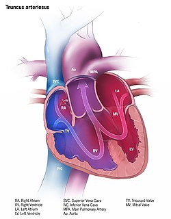
Persistent truncus arteriosus (PTA), often referred to simply as Truncus Arteriosus, is a rare form of congenital heart disease that presents at birth. In this condition, the embryological structure known as the truncus arteriosus fails to properly divide into the pulmonary trunk and aorta. This results in one arterial trunk arising from the heart and providing mixed blood to the coronary arteries, pulmonary arteries, and systemic circulation. For the International Classification of Diseases (ICD-11), the International Paediatric and Congenital Cardiac Code (IPCCC) was developed to standardize the nomenclature of congenital heart disease. Under this system, English is now the official language, and persistent truncus arteriosus should properly be termed Common arterial trunk.

The bulbus glandis is an erectile tissue structure on the penis of canid mammals. During mating, immediately before ejaculation the tissues swell up to lock (tie) the male's penis inside the female. The locking is completed by circular muscles just inside the female's vagina; this is called "the knot" tightening thus preventing the male from withdrawing. The circular muscles also contract intermittently, which has the effect of stimulating ejaculation of sperm, followed by prostatic fluid, as well as maintaining the swelling of the penis and therefore the tie, for some time. For domestic dogs the tie may last up to half an hour or more, though usually less. When male canines are excited, the bulbus glandis may swell up inside the penile sheath, even if the dog has been neutered.

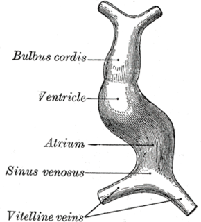
The bulbus cordis is a part of the developing heart that lies ventral to the primitive ventricle after the heart assumes its S-shaped form. The superior end of the bulbus cordis is also called the conotruncus.
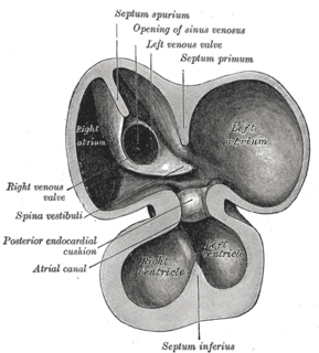
The sinus venosus is a large quadrangular cavity which precedes the atrium on the venous side of the chordate heart. In mammals, it exists distinctly only in the embryonic heart ; however, the sinus venosus persists in the adult. In the adult, it is incorporated into the wall of the right atrium to form a smooth part called the sinus venarum, which is separated from the rest of the atrium by a ridge of fibres called the crista terminalis. The sinus venosus also forms the SA node and the coronary sinus; in (most) mammals only.

The infundibulum is a conical pouch formed from the upper and left angle of the right ventricle in the chordate heart, from which the pulmonary trunk arises. It develops from the bulbus cordis. Typically, the infundibulum refers to the corresponding internal structure, whereas the conus arteriosus refers to the external structure. Defects in infundibulum development can result in a heart condition known as tetralogy of Fallot.

The primitive ventricle or embryonic ventricle of the developing heart, together with the bulbus cordis that lies in front of it, gives rise to the left and right ventricles. The primitive ventricle provides the trabeculated parts of the walls, and the bulbus cordis the smooth parts.
The primitive atrium is a stage in the embryonic development of the human heart. It grows rapidly and partially encircles the bulbus cordis; the groove against which the bulbus cordis lies is the first indication of a division into right and left atria.

The long posterior ciliary arteries are arteries of the head arising, together with the other ciliary arteries, from the ophthalmic artery. There are two in each eye.
A ventricular outflow tract is a portion of either the left ventricle or right ventricle of the heart through which blood passes in order to enter the great arteries.

The aorticopulmonary septum is developmentally formed from neural crest, specifically the cardiac neural crest, and actively separates the aorta and pulmonary arteries and fuses with the interventricular septum within the heart during heart development.
The truncus arteriosus is a structure that is present during embryonic development. It is an arterial trunk that originates from both ventricles of the heart that later divides into the aorta and the pulmonary trunk.
The heart is the first functional organ in a vertebrate embryo. There are 5 stages to heart development.
The Zebrafish Information Network is an online biological database of information about the zebrafish. The zebrafish is a widely used model organism for genetic, genomic, and developmental studies, and ZFIN provides an integrated interface for querying and displaying the large volume of data generated by this research. To facilitate use of the zebrafish as a model of human biology, ZFIN links these data to corresponding information about other model organisms and to human disease databases. Abundant links to external sequence databases and to genome browsers are included. Gene product, gene expression, and phenotype data are annotated with terms from biomedical ontologies. ZFIN is based at the University of Oregon in the United States, with funding provided by the National Institutes of Health (NIH).
The tubular heart or primitive heart tube is the earliest stage of heart development.

The posterior auricular muscle consists of two or three fleshy fasciculi, which arise from the mastoid portion of the temporal bone by short aponeurotic fibers. They are inserted into the lower part of the cranial surface of the concha.
Bulbus may refer to:

Heart development refers to the prenatal development of the heart. This begins with the formation of two endocardial tubes which merge to form the tubular heart, also called the primitive heart tube. The heart is the first functional organ in vertebrate embryos, and in the human, beats spontaneously around week 5 of development.
References
1. "ZFIN: Anatomical Structure: Bulbus Arteriosus." ZFIN: The Zebrafish Model Organism Database. Web. 8 May 2011. <http://zfin.org/action/anatomy/term-detail?anatomyItem.zdbID=ZDB-ANAT-011113-107>.
| | This fish-related article is a stub. You can help Wikipedia by expanding it. |