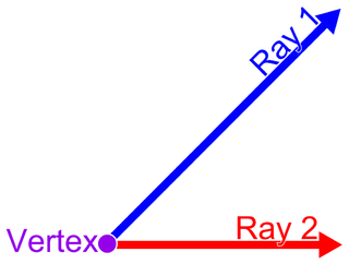
In Euclidean geometry, an angle is the figure formed by two rays, called the sides of the angle, sharing a common endpoint, called the vertex of the angle. Angles formed by two rays lie in the plane that contains the rays. Angles are also formed by the intersection of two planes. These are called dihedral angles. Two intersecting curves may also define an angle, which is the angle of the rays lying tangent to the respective curves at their point of intersection.

Electrocardiography is the process of producing an electrocardiogram, a recording of the heart's electrical activity. It is an electrogram of the heart which is a graph of voltage versus time of the electrical activity of the heart using electrodes placed on the skin. These electrodes detect the small electrical changes that are a consequence of cardiac muscle depolarization followed by repolarization during each cardiac cycle (heartbeat). Changes in the normal ECG pattern occur in numerous cardiac abnormalities, including cardiac rhythm disturbances, inadequate coronary artery blood flow, and electrolyte disturbances.

Willem Einthoven was a Dutch doctor and physiologist. He invented the first practical electrocardiograph in 1895 and received the Nobel Prize in Physiology or Medicine in 1924 for it.
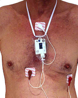
In medicine, a Holter monitor is a type of ambulatory electrocardiography device, a portable device for cardiac monitoring for at least 24 hours.

Dextrocardia is a rare congenital condition in which the apex of the heart is located on the right side of the body. There are two main types of dextrocardia: dextrocardia of embryonic arrest and dextrocardia situs inversus. Dextrocardia situs inversus is further divided.
The QRS complex is the combination of three of the graphical deflections seen on a typical electrocardiogram. It is usually the central and most visually obvious part of the tracing. It corresponds to the depolarization of the right and left ventricles of the heart and contraction of the large ventricular muscles.

Left bundle branch block (LBBB) is a cardiac conduction abnormality seen on the electrocardiogram (ECG). In this condition, activation of the left ventricle of the heart is delayed, which causes the left ventricle to contract later than the right ventricle.

Motion, the process of movement, is described using specific anatomical terms. Motion includes movement of organs, joints, limbs, and specific sections of the body. The terminology used describes this motion according to its direction relative to the anatomical position of the body parts involved. Anatomists and others use a unified set of terms to describe most of the movements, although other, more specialized terms are necessary for describing unique movements such as those of the hands, feet, and eyes.

A string galvanometer is a sensitive fast-responding measuring instrument that uses a single fine filament of wire suspended in a strong magnetic field to measure small currents. In use, a strong light source is used to illuminate the fine filament, and the optical system magnifies the movement of the filament allowing it to be observed or recorded by photography. The principle of the string galvanometer remained in use for electrocardiograms until the advent of electronic vacuum-tube amplifiers in the 1920s.

The hexaxial reference system, better known as the Cabrera system, is a convention to present the extremity leads of the 12 lead electrocardiogram, that provides an illustrative logical sequence that helps interpretation of the ECG, especially to determine the heart's electrical axis in the frontal plane. The most practical way of using this is by arranging extremity leads according to the Cabrera system, reversing polarity of lead aVR and presenting ECG complexes in the order. Then determine the direction the maximal ECG vector is "pointing", i.e. in which lead there are most positive amplitude - this direction is the electrical axis - see diagram. Example: If lead I has the highest amplitude, the axis is approximately 0°. Conversely, if lead III has the most negative amplitude it means the vector is pointing away from this lead, i.e. towards -60°.
The electrical axis of the heart is the net direction in which the wave of depolarization travels. It is measured using an electrocardiogram (ECG). Normally, this begins at the sinoatrial node ; from here the wave of depolarisation travels down to the apex of the heart. The hexaxial reference system can be used to visualise the directions in which the depolarisation wave may travel.
Left anterior fascicular block (LAFB) is an abnormal condition of the left ventricle of the heart, related to, but distinguished from, left bundle branch block (LBBB).
A Bioamplifier is an electrophysiological device, a variation of the instrumentation amplifier, used to gather and increase the signal integrity of physiologic electrical activity for output to various sources. It may be an independent unit, or integrated into the electrodes.
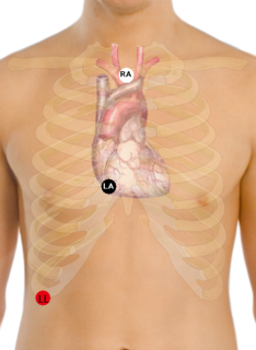
A Lewis Lead is a modified ECG lead used to detect atrial flutter waves when atrial flutter is suspected clinically, based on signs and symptoms, but is not definitely demonstrated on the standard 12 lead ECG. In order to create the Lewis Lead, the right arm electrode is moved to the manubrium adjacent to the sternum. Then the left arm electrode is moved to the right, fifth intercostal space adjacent to the sternum. The left leg electrode is placed on the right lower costal margin. The Lewis Lead is then read as Lead I on the ECG and, since in most patients it will be roughly perpendicular to the wave of ventricular depolarization, atrial flutter waves may be more apparent.

Vectorcardiography (VCG) is a method of recording the magnitude and direction of the electrical forces that are generated by the heart by means of a continuous series of vectors that form curving lines around a central point.

ST elevation refers to a finding on an electrocardiogram wherein the trace in the ST segment is abnormally high above the baseline.
In electrocardiography, left axis deviation (LAD) is a condition wherein the mean electrical axis of ventricular contraction of the heart lies in a frontal plane direction between −30° and −90°. This is reflected by a QRS complex positive in lead I and negative in leads aVF and II.

In mathematics, a unit circle is a circle of unit radius—that is, a radius of 1. Frequently, especially in trigonometry, the unit circle is the circle of radius 1 centered at the origin in the Cartesian coordinate system in the Euclidean plane. In topology, it is often denoted as S1 because it is a one-dimensional unit n-sphere.
Wireless ambulatory electrocardiography (ECG) is a type of ambulatory electrocardiography with recording devices that use wireless technology, such as Bluetooth and smartphones, for at-home cardiac monitoring. These devices are generally recommended to people who have been previously diagnosed with arrhythmias and want to have them monitored, or for those who have suspected arrhythmias and need to be monitored over an extended period of time in order to be diagnosed.

The forward problem of electrocardiology is a computational and mathematical approach to study the electrical activity of the heart through the body surface. The principal aim of this study is to computationally reproduce an electrocardiogram (ECG), which has important clinical relevance to define cardiac pathologies such as ischemia and infarction, or to test pharmaceutical intervention. Given their important functionalities and the relative small invasiveness, the electrocardiography techniques are used quite often as clinical diagnostic tests. Thus, it is natural to proceed to computationally reproduce an ECG, which means to mathematically model the cardiac behaviour inside the body.


