Fourier-transform spectroscopy is a measurement technique whereby spectra are collected based on measurements of the coherence of a radiative source, using time-domain or space-domain measurements of the electromagnetic radiation or other type of radiation. It can be applied to a variety of types of spectroscopy including optical spectroscopy, infrared spectroscopy, nuclear magnetic resonance (NMR) and magnetic resonance spectroscopic imaging (MRSI), mass spectrometry and electron spin resonance spectroscopy. There are several methods for measuring the temporal coherence of the light, including the continuous wave Michelson or Fourier-transform spectrometer and the pulsed Fourier-transform spectrograph.
Cognitive neuroscience is the scientific field that is concerned with the study of the biological processes and aspects that underlie cognition, with a specific focus on the neural connections in the brain which are involved in mental processes. It addresses the questions of how cognitive activities are affected or controlled by neural circuits in the brain. Cognitive neuroscience is a branch of both neuroscience and psychology, overlapping with disciplines such as behavioral neuroscience, cognitive psychology, physiological psychology and affective neuroscience. Cognitive neuroscience relies upon theories in cognitive science coupled with evidence from neurobiology, and computational modeling.

Functional magnetic resonance imaging or functional MRI (fMRI) measures brain activity by detecting changes associated with blood flow. This technique relies on the fact that cerebral blood flow and neuronal activation are coupled. When an area of the brain is in use, blood flow to that region also increases.

Functional neuroimaging is the use of neuroimaging technology to measure an aspect of brain function, often with a view to understanding the relationship between activity in certain brain areas and specific mental functions. It is primarily used as a research tool in cognitive neuroscience, cognitive psychology, neuropsychology, and social neuroscience.

An event-related potential (ERP) is the measured brain response that is the direct result of a specific sensory, cognitive, or motor event. More formally, it is any stereotyped electrophysiological response to a stimulus. The study of the brain in this way provides a noninvasive means of evaluating brain functioning.
Statistical parametric mapping or SPM is a statistical technique for examining differences in brain activity recorded during functional neuroimaging experiments. It was created by Karl Friston It may alternatively refer to software created by the Wellcome Department of Imaging Neuroscience to carry out such analyses.
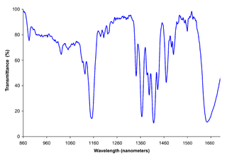
Near-infrared spectroscopy (NIRS) is a spectroscopic method that uses the near-infrared region of the electromagnetic spectrum. Typical applications include medical and physiological diagnostics and research including blood sugar, pulse oximetry, functional neuroimaging, sports medicine, elite sports training, ergonomics, rehabilitation, neonatal research, brain computer interface, urology, and neurology. There are also applications in other areas as well such as pharmaceutical, food and agrochemical quality control, atmospheric chemistry, combustion research and astronomy.
Hemoencephalography (HEG) is a relatively new neurofeedback technique within the field of neurotherapy. Neurofeedback, a specific form of biofeedback, is based on the idea that human beings can consciously alter their brain function through training sessions in which they attempt to change the signal generated by their brain and measured via some neurological feedback mechanism. By so doing, participants increase cerebral blood flow to a specified region of the brain, consequently increasing brain activity and performance on tasks involving that region of the brain.
Medical optical imaging is the use of light as an investigational imaging technique for medical applications. Examples include optical microscopy, spectroscopy, endoscopy, scanning laser ophthalmoscopy, and optical coherence tomography. Because light is an electromagnetic wave, similar phenomena occur in X-rays, microwaves, and radio waves.

Functional Near-Infrared Spectroscopy (fNIRS), is the use of near-infrared spectroscopy (NIRS) for the purpose of functional neuroimaging. Using fNIRS, brain activity is measured through hemodynamic responses associated with neuron behaviour.

Molecular imaging originated from the field of radiopharmacology due to the need to better understand fundamental molecular pathways inside organisms in a noninvasive manner.

Optical tomography is a form of computed tomography that creates a digital volumetric model of an object by reconstructing images made from light transmitted and scattered through an object. Optical tomography is used mostly in medical imaging research. Optical tomography in industry is used as a sensor of thickness and internal structure of semiconductors.
Chemical imaging is the analytical capability to create a visual image of components distribution from simultaneous measurement of spectra and spatial, time information. Hyperspectral imaging measures contiguous spectral bands, as opposed to multispectral imaging which measures spaced spectral bands.
Arno Villringer is a director at the Max Planck Institute for Human Cognitive and Brain Sciences in Leipzig, Germany; director of the Department of Cognitive Neurology at University Hospital Leipzig; and Academic Director of the Berlin School of Mind and Brain and the Mind&Brain Institute, Berlin. He holds a full professorship at University of Leipzig and an honorary professorship at Charité, Humboldt-Universität zu Berlin.
Neuroimaging intelligence testing concerns the use of neuroimaging techniques to evaluate human intelligence. Neuroimaging technology has advanced such that scientists hope to use neuroimaging increasingly for investigations of brain function related to IQ.

Resting state fMRI is a method of functional magnetic resonance imaging (fMRI) that is used in brain mapping to evaluate regional interactions that occur in a resting or task-negative state, when an explicit task is not being performed. A number of resting-state conditions are identified in the brain, one of which is the default mode network. These resting brain state conditions are observed through changes in blood flow in the brain which creates what is referred to as a blood-oxygen-level dependent (BOLD) signal that can be measured using fMRI. Because brain activity is intrinsic, present even in the absence of an externally prompted task, any brain region will have spontaneous fluctuations in BOLD signal. The resting state approach is useful to explore the brain's functional organization and to examine if it is altered in neurological or mental disorders. Resting-state functional connectivity research has revealed a number of networks which are consistently found in healthy subjects, different stages of consciousness and across species, and represent specific patterns of synchronous activity.

Alcohol-related brain damage alters both the structure and function of the brain as a result of the direct neurotoxic effects of alcohol intoxication or acute alcohol withdrawal. Increased alcohol intake is associated with damage to brain regions including the frontal lobe, limbic system, and cerebellum, with widespread cerebral atrophy, or brain shrinkage caused by neuron degeneration. This damage can be seen on neuroimaging scans.
Neal J. Cohen is a professor of psychology in the Cognitive Neuroscience division of the University of Illinois at Urbana–Champaign. He is appointed as a full-time faculty member in the Beckman Institute for Advanced Science and Technology at the University of Illinois. He is the founding director of the Center for Nutrition, Learning, and Memory (CNLM), a partnership of the University of Illinois and Abbott Laboratories as of 2011. He is also the founding director of the Interdisciplinary Health Sciences Initiative (IHSI) at the University of Illinois, formed 2014.
Time-domain diffuse optics or time-resolved functional near-infrared spectroscopy is branch of functional near-Infrared spectroscopy which deals with light propagation in diffusive media. There are three main approaches to diffuse optics namely continuous wave (CW), frequency domain (FD) and time-domain (TD). Biological tissue in the range of red to near-infrared wavelengths are transparent to light and can be used to probe deep layers of the tissue thus enabling various in vivo applications and clinical trials.


