
The vestibulo-ocular reflex (VOR) is a reflex that acts to stabilize gaze during head movement, with eye movement due to activation of the vestibular system, it is also known as the Cervico-ocular reflex. The reflex acts to stabilize images on the retinas of the eye during head movement. Gaze is held steadily on a location by producing eye movements in the direction opposite that of head movement. For example, when the head moves to the right, the eyes move to the left, meaning the image a person sees stays the same even though the head has turned. Since slight head movement is present all the time, VOR is necessary for stabilizing vision: people with an impaired reflex find it difficult to read using print, because the eyes do not stabilise during small head tremors, and also because damage to reflex can cause nystagmus.

The vestibular system, in vertebrates, is a sensory system that creates the sense of balance and spatial orientation for the purpose of coordinating movement with balance. Together with the cochlea, a part of the auditory system, it constitutes the labyrinth of the inner ear in most mammals.
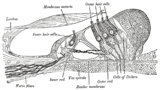
Hair cells are the sensory receptors of both the auditory system and the vestibular system in the ears of all vertebrates, and in the lateral line organ of fishes. Through mechanotransduction, hair cells detect movement in their environment.
In neuroscience, parvocellular cells, also called P-cells, are neurons located within the parvocellular layers of the lateral geniculate nucleus (LGN) of the thalamus. Their name comes from Latin parvus 'small', due to the small size of the cell compared to the larger magnocellular cells. Phylogenetically, parvocellular neurons are more modern than magnocellular ones.
In animals, including humans, the startle response is a largely unconscious defensive response to sudden or threatening stimuli, such as sudden noise or sharp movement, and is associated with negative affect. Usually the onset of the startle response is a startle reflex reaction. The startle reflex is a brainstem reflectory reaction (reflex) that serves to protect vulnerable parts, such as the back of the neck and the eyes (eyeblink) and facilitates escape from sudden stimuli. It is found across many different species, throughout all stages of life. A variety of responses may occur depending on the affected individual's emotional state, body posture, preparation for execution of a motor task, or other activities. The startle response is implicated in the formation of specific phobias.

Excitatory amino-acid transporter 4 (EAAT4) is a protein that in humans is encoded by the SLC1A6 gene.

The Kir2.1 inward-rectifier potassium channel is a lipid-gated ion channel encoded by the KCNJ2 gene.

Neuronal acetylcholine receptor subunit beta-4 is a protein that in humans is encoded by the CHRNB4 gene.
Voltage-dependent L-type calcium channel subunit beta-2 is a protein that in humans is encoded by the CACNB2 gene.

Monocarboxylate transporter 5 is a protein that in humans is encoded by the SLC16A4 gene.

Potassium inwardly-rectifying channel, subfamily J, member 15, also known as KCNJ15 is a human gene, which encodes the Kir4.2 protein.

Sodium–hydrogen exchanger 2 is a protein that in humans is encoded by the SLC9A2 gene.
Chloride channel accessory 2 is a protein that in humans is encoded by the CLCA2 gene.

Neuronal acetylcholine receptor subunit beta-3 is a protein that in humans is encoded by the CHRNB3 gene. This gene has been identified as a candidate for predisposition to tobacco dependence.

Potassium inwardly-rectifying channel, subfamily J, member 16 (KCNJ16) is a human gene encoding the Kir5.1 protein.
The vestibular evoked myogenic potential is a neurophysiological assessment technique used to determine the function of the otolithic organs of the inner ear. It complements the information provided by caloric testing and other forms of inner ear testing. There are two different types of VEMPs. One is the oVEMP and another is the cVEMP. The oVEMP measures integrity of the utricule and superior vestibular nerve and the cVemp measures the saccule and the inferior vestibular nerve.

Myosin-4 also known as myosin, heavy chain 4 is a protein which in humans is encoded by the MYH4 gene.
The righting reflex, also known as the labyrinthine righting reflex, or the Cervico-collic reflex; is a reflex that corrects the orientation of the body when it is taken out of its normal upright position. It is initiated by the vestibular system, which detects that the body is not erect and causes the head to move back into position as the rest of the body follows. The perception of head movement involves the body sensing linear acceleration or the force of gravity through the otoliths, and angular acceleration through the semicircular canals. The reflex uses a combination of visual system inputs, vestibular inputs, and somatosensory inputs to make postural adjustments when the body becomes displaced from its normal vertical position. These inputs are used to create what is called an efference copy. This means that the brain makes comparisons in the cerebellum between expected posture and perceived posture, and corrects for the difference. The reflex takes 6 or 7 weeks to perfect, but can be affected by various types of balance disorders.
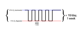
Intermittent hypoxia (also known as episodic hypoxia) is an intervention in which a person or animal undergoes alternating periods of normoxia and hypoxia. Normoxia is defined as exposure to oxygen levels normally found in Earth's atmosphere (~21% O2) and hypoxia as any oxygen levels lower than those of normoxia. Normally, exposure to hypoxia is negatively associated to physiological changes to the body, such as altitude sickness. However, when used in moderation, intermittent hypoxia may be used clinically as a means to alleviate various pathological conditions.
Gait variability seen in Parkinson's Disorders arise due to cortical changes induced by pathophysiology of the disease process. Gait rehabilitation is focused to harness the adapted connections involved actively to control these variations during the disease progression. Gait variabilities seen are attributed to the defective inputs from the Basal Ganglia. However, there is altered activation of other cortical areas that support the deficient control to bring about a movement and maintain some functional mobility.