Carbachol, also known as carbamylcholine, is a cholinomimetic drug that binds and activates acetylcholine receptors. Thus it is classified as a cholinergic agonist. It is primarily used for various ophthalmic purposes, such as for treating glaucoma, or for use during ophthalmic surgery. It is generally administered as an ophthalmic solution.

The reticular formation is a set of interconnected nuclei that are located throughout the brainstem. The reticular formation is not anatomically well defined because it includes neurons located in different parts of the brain. The neurons of the reticular formation make up a complex set of networks in the core of the brainstem that extend from the upper part of the midbrain to the lower part of the medulla oblongata. The reticular formation includes ascending pathways to the cortex in the ascending reticular activating system (ARAS) and descending pathways to the spinal cord via the reticulospinal tracts of the descending reticular formation.

The pontine tegmentum, or dorsal pons, is located within the brainstem, and is one of two parts of the pons, the other being the ventral pons or basilar part of the pons. The pontine tegmentum can be defined in contrast to the basilar pons: basilar pons contains the corticospinal tract running craniocaudally and can be considered the rostral extension of the ventral medulla oblongata; however, basilar pons is distinguished from ventral medulla oblongata in that it contains additional transverse pontine fibres that continue laterally to become the middle cerebellar peduncle. The pontine tegmentum is all the material dorsal from the basilar pons to the fourth ventricle. Along with the dorsal surface of the medulla, it forms part of the rhomboid fossa – the floor of the fourth ventricle.

The ventrolateral preoptic nucleus (VLPO), also known as the intermediate nucleus of the preoptic area (IPA), is a small cluster of neurons situated in the anterior hypothalamus, sitting just above and to the side of the optic chiasm in the brain of humans and other animals. The brain's sleep-promoting nuclei, together with the ascending reticular activating system and the widely-projecting system of orexin neurons in the lateral hypothalamus, are the interconnected neural systems which control states of arousal, sleep, and transitions between these two states. The VLPO is active during sleep, primarily during non-rapid eye movement sleep, and releases inhibitory neurotransmitters, mainly GABA and galanin, which inhibit neurons of the ascending reticular activating system that are involved in wakefulness and arousal. The VLPO is in turn innervated by neurons from the aforementioned neural systems. The VLPO is activated by the sleep-inducing neurotransmitters serotonin and adenosine and endosomnogen Prostaglandin D2. The VLPO is inhibited during wakefulness by the arousal-inducing neurotransmitters norepinephrine and acetylcholine. The role of the VLPO in sleep and wakefulness, and its association with sleep disorders – particularly insomnia and narcolepsy – is a growing area of neuroscience research.

The paramedian pontine reticular formation, also known as PPRF or paraabducens nucleus, is part of the pontine reticular formation, a brain region without clearly defined borders in the center of the pons. It is involved in the coordination of eye movements, particularly horizontal gaze and saccades.
The reticulotegmental nucleus, tegmental pontine reticular nucleus is an area within the floor of the midbrain. This area is known to affect the cerebellum with its axonal projections.
The parvocellular reticular nucleus is part of the brain located dorsolateral to the caudal pontine reticular nucleus.
The oral pontine reticular nucleus, or rostral pontine reticular nucleus, is delineated from the caudal pontine reticular nucleus. This nucleus tapers into the lower mesencephalic reticular formation and contains sporadic giant cells.
The caudal pontine reticular nucleus or nucleus reticularis pontis caudalis is composed of gigantocellular neurons.
The gigantocellular nucleus is a subregion of the medullary reticular formation. As the name indicates, is mainly composed of the so-called giant neuronal cells.
The paramedian reticular nucleus sends its connections to the spinal cord in a mostly ipsilateral manner, although there is some decussation.
Ponto-geniculo-occipital waves or PGO waves are phasic field potentials. These waves can be recorded from the pons, the lateral geniculate nucleus (LGN), and the occipital cortex regions of the brain, where these waveforms originate. The waves begin as electrical pulses from the pons, then move to the lateral geniculate nucleus residing in the thalamus, and then finally end up in the primary visual cortex of the occipital lobe. The appearances of these waves are most prominent in the period right before rapid eye movement sleep, and are theorized to be intricately involved with eye movement of both wake and sleep cycles in many different animals.
The midbrain reticular formation(MRF) also reticular formation of midbrain, mesencephalic reticular formation, tegmental reticular formation, formatio reticularis (tegmenti) mesencephali) is a structure in the midbrain consisting of the dorsal tegmental nucleus, ventral tegmental nucleus, and cuneiform nucleus. These are also known as the tegmental nuclei.
The activation-synthesis hypothesis, proposed by Harvard University psychiatrists John Allan Hobson and Robert McCarley, is a neurobiological theory of dreams first published in the American Journal of Psychiatry in December 1977. The differences in neuronal activity of the brainstem during waking and REM sleep were observed, and the hypothesis proposes that dreams result from brain activation during REM sleep. Since then, the hypothesis has undergone an evolution as technology and experimental equipment has become more precise. Currently, a three-dimensional model called AIM Model, described below, is used to determine the different states of the brain over the course of the day and night. The AIM Model introduces a new hypothesis that primary consciousness is an important building block on which secondary consciousness is constructed.

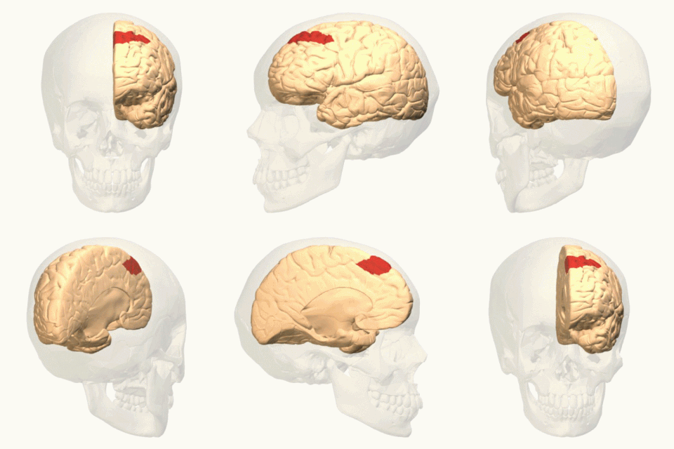
In neuroanatomy, corticomesencephalic tract is a descending nerve tract that originates in the frontal eye field and terminate in the midbrain. Its fibers mediate conjugate eye movement.
Reticular nucleus may refer to:
The parafacial zone (PZ) is a brain structure located in the brain stem within the medulla oblongata. Due to a high presence of GABAergic neurons, as well as its involvement in slow-wave sleep, the parafacial zone is believed to be heavily responsible for non-rapid eye movement (non-REM) sleep regulation.


