
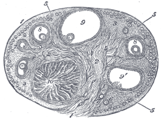
The ovary is an organ found in the female reproductive system that produces an ovum. When released, this travels down the fallopian tube into the uterus, where it may become fertilized by a sperm. There is an ovary found on the left and right sides of the body. The ovaries also secrete hormones that play a role in the menstrual cycle and fertility. The ovary progresses through many stages beginning in the prenatal period through menopause. It is also an endocrine gland because of the various hormones that it secretes.

The chorion is the outermost fetal membrane around the embryo in mammals, birds and reptiles. It develops from an outer fold on the surface of the yolk sac, which lies outside the zona pellucida, known as the vitelline membrane in other animals. In insects it is developed by the follicle cells while the egg is in the ovary.
The corpus luteum is a temporary endocrine structure in female ovaries and is involved in the production of relatively high levels of progesterone, moderate levels of estradiol and inhibin A, and small amounts of estrogen. It is the remains of the ovarian follicle that has released a mature ovum during a previous ovulation.

An ovarian follicle is a roughly spheroid cellular aggregation set found in the ovaries. It secretes hormones that influence stages of the menstrual cycle. Women begin puberty with about 400,000 follicles, each with the potential to release an egg cell (ovum) at ovulation for fertilization. These eggs are developed once every menstrual cycle.

The corpus albicans is the regressed form of the corpus luteum. As the corpus luteum is being broken down by macrophages, fibroblasts lay down type I collagen, forming the corpus albicans. This process is called "luteolysis". The remains of the corpus albicans may persist as a scar on the surface of the ovary.

The zona pellucida is a glycoprotein layer surrounding the plasma membrane of mammalian oocytes. It is a vital constitutive part of the oocyte. The zona pellucida first appears in unilaminar primary oocytes. It is secreted by both the oocyte and the ovarian follicles. The zona pellucida is surrounded by the cumulus oophorus. The cumulus is composed of cells that care for the egg when it is emitted from the ovary.

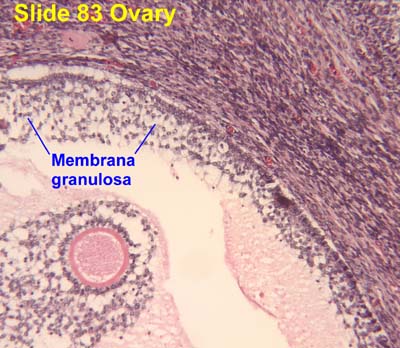
A granulosa cell or follicular cell is a somatic cell of the sex cord that is closely associated with the developing female gamete in the ovary of mammals.

In biology, folliculogenesis is the maturation of the ovarian follicle, a densely packed shell of somatic cells that contains an immature oocyte. Folliculogenesis describes the progression of a number of small primordial follicles into large preovulatory follicles that occurs in part during the menstrual cycle.

The tunica media, or media for short, is the middle tunica (layer) of an artery or vein. It lies between the tunica intima on the inside and the tunica externa on the outside.
The corona radiata is the innermost layer of the cells of the cumulus oophorus and is directly adjacent to the zona pellucida, the inner protective glycoprotein layer of the ovum. Its main purpose in many animals is to supply vital proteins to the cell. It is formed by follicle cells adhering to the oocyte before it leaves the ovarian follicle, and originates from the squamous granulosa cells present at the primordial stage of follicular development. The corona radiata is formed when the granulosa cells enlarge and become cuboidal, which occurs during the transition from the primordial to primary stage. These cuboidal granulosa cells, also known as the granulosa radiata, form more layers throughout the maturation process, and remain attached to the zona pellucida after the ovulation of the Graafian follicle. For fertilization to occur, sperm cells rely on hyaluronidase to disperse the corona radiata from the zona pellucida of the secondary (ovulated) oocyte, thus permitting entry into the perivitelline space and allowing contact between the sperm cell and the nucleus of the oocyte.
Theca interna cells express receptors for luteinizing hormone (LH) to produce androstenedione, which via a few steps, gives the granulosa the precursor for estrogen manufacturing.

The outer nuclear layer, is one of the layers of the vertebrate retina, the light-detecting portion of the eye. Like the inner nuclear layer, the outer nuclear layer contains several strata of oval nuclear bodies; they are of two kinds, viz.: rod and cone granules, so named on account of their being respectively connected with the rods and cones of the next layer.

In the female reproductive system, the fimbria is a fringe of tissue around the ostium of the Fallopian tube, in the direction of the ovary.
The medulla of ovary is a highly vascular stroma in the center of the ovary. It forms from embryonic mesenchyme and contains blood vessels, lymphatic vessels, and nerves.
The ovarian surface epithelium, also called the germinal epithelium of Waldeyer, is a layer of simple squamous-to-cuboidal epithelial cells covering the ovary.

The germinal epithelium is the innermost layer of the testicle.
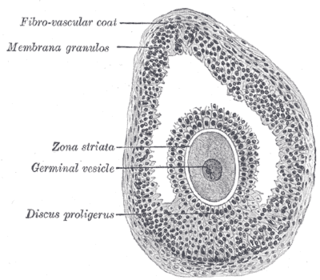
The cumulus oophorus, also called discus proligerus, is a cluster of cells that surround the oocyte both in the ovarian follicle and after ovulation. In the antral follicle, it may be regarded as an extension of the membrana granulosa. The innermost layer of these cells is the corona radiata.
The theca folliculi comprise a layer of the ovarian follicles. They appear as the follicles become secondary follicles.
Before the fertilized ovum reaches the uterus, the mucous membrane of the body of the uterus undergoes important changes and is then known as the decidua. The thickness and vascularity of the mucous membrane are greatly increased; its glands are elongated and open on its free surface by funnel-shaped orifices, while their deeper portions are tortuous and dilated into irregular spaces. The interglandular tissue is also increased in quantity, and is crowded with large round, oval, or polygonal cells, termed decidual cells. Their enlargement is due to glycogen and lipid accumulation in the cytoplasm allowing these cells to provide a rich source of nutrition for the developing embryo.
The theca externa is the outer layer of the theca folliculi. It is derived from connective tissue, the cells resembling fibroblasts, and contains abundant collagen. During ovulation, the surge in luteinizing hormone increases cAMP which increases progesterone and PGF2α production. The PGF2α induces the contraction of the smooth muscle cells of the theca externa, increasing intrafollicular pressure. This aids in rupture of the mature oocyte, or immature oocyte at the germinal vesicle stage in the canine, along with plasmin and collagenase degradation of the follicle wall.

