Related Research Articles

A bone is a rigid tissue that constitutes part of the vertebrate skeleton in animals. Bones protect the various organs of the body, produce red and white blood cells, store minerals, provide structure and support for the body, and enable mobility. Bones come in a variety of shapes and sizes and have a complex internal and external structure. They are lightweight yet strong and hard, and serve multiple functions.

The middle ear is the portion of the ear internal to the eardrum, and external to the oval window of the inner ear. The mammalian middle ear contains three ossicles, which transfer the vibrations of the eardrum into waves in the fluid and membranes of the inner ear. The hollow space of the middle ear is also known as the tympanic cavity and is surrounded by the tympanic part of the temporal bone. The auditory tube joins the tympanic cavity with the nasal cavity (nasopharynx), allowing pressure to equalize between the middle ear and throat.

Soft tissue is all the tissue in the body that is not hardened by the processes of ossification or calcification such as bones and teeth. Soft tissue connects, surrounds or supports internal organs and bones, and includes muscle, tendons, ligaments, fat, fibrous tissue, skin, lymph and blood vessels, fasciae, and synovial membranes.

The fibula or calf bone is a leg bone on the lateral side of the tibia, to which it is connected above and below. It is the smaller of the two bones and, in proportion to its length, the slenderest of all the long bones. Its upper extremity is small, placed toward the back of the head of the tibia, below the knee joint and excluded from the formation of this joint. Its lower extremity inclines a little forward, so as to be on a plane anterior to that of the upper end; it projects below the tibia and forms the lateral part of the ankle joint.

The long bones are those that are longer than they are wide. They are one of five types of bones: long, short, flat, irregular and sesamoid. Long bones, especially the femur and tibia, are subjected to most of the load during daily activities and they are crucial for skeletal mobility. They grow primarily by elongation of the diaphysis, with an epiphysis at each end of the growing bone. The ends of epiphyses are covered with hyaline cartilage. The longitudinal growth of long bones is a result of endochondral ossification at the epiphyseal plate. Bone growth in length is stimulated by the production of growth hormone (GH), a secretion of the anterior lobe of the pituitary gland.

A bone fracture is a medical condition in which there is a partial or complete break in the continuity of the bone. In more severe cases, the bone may be broken into several pieces. A bone fracture may be the result of high force impact or stress, or a minimal trauma injury as a result of certain medical conditions that weaken the bones, such as osteoporosis, osteopenia, bone cancer, or osteogenesis imperfecta, where the fracture is then properly termed a pathologic fracture.

A trabecula is a small, often microscopic, tissue element in the form of a small beam, strut or rod that supports or anchors a framework of parts within a body or organ. A trabecula generally has a mechanical function, and is usually composed of dense collagenous tissue. They can be composed of other materials such as muscle and bone. In the heart, muscles form trabeculae carneae and septomarginal trabecula. Cancellous bone is formed from groupings of trabeculated bone tissue.

A dental implant is a surgical component that interfaces with the bone of the jaw or skull to support a dental prosthesis such as a crown, bridge, denture, facial prosthesis or to act as an orthodontic anchor. The basis for modern dental implants is a biologic process called osseointegration, in which materials such as titanium form an intimate bond to bone. The implant fixture is first placed so that it is likely to osseointegrate, then a dental prosthetic is added. A variable amount of healing time is required for osseointegration before either the dental prosthetic is attached to the implant or an abutment is placed which will hold a dental prosthetic/crown.
Osseointegration is the direct structural and functional connection between living bone and the surface of a load-bearing artificial implant. A more recent definition defines osseointegration as "functional ankylosis ", where new bone is laid down directly on the implant surface and the implant exhibits mechanical stability. Osseointegration has enhanced the science of medical bone and joint replacement techniques as well as dental implants and improving prosthetics for amputees.
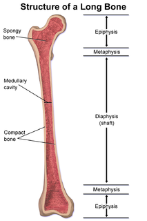
The metaphysis is the neck portion of a long bone between the epiphysis and the diaphysis. It contains the growth plate, the part of the bone that grows during childhood, and as it grows it ossifies near the diaphysis and the epiphyses. The metaphysis contains a diverse population of cells including mesenchymal stem cells, which give rise to bone and fat cells, as well as hematopoietic stem cells which give rise to a variety of blood cells as well as bone-destroying cells called osteoclasts. Thus the metaphysis contains a highly metabolic set of tissues including trabecular (spongy) bone, blood vessels, as well as Marrow Adipose Tissue (MAT).

The periodontal ligament, commonly abbreviated as the PDL, is a group of specialized connective tissue fibers that essentially attach a tooth to the alveolar bone within which it sits. It inserts into root cementum one side and onto alveolar bone on the other.

The brow ridge, or supraorbital ridge known as superciliary arch in medicine, refers to a bony ridge located above the eye sockets of all primates. In Homo sapiens sapiens the eyebrows are located on their lower margin.
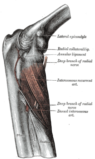
The interosseous membrane of the forearm is a fibrous sheet that connects the interosseous margins of the radius and the ulna. It is the main part of the radio-ulnar syndesmosis, a fibrous joint between the two bones.
The Mechanostat is a term describing the way in which mechanical loading influences bone structure by changing the mass and architecture to provide a structure that resists habitual loads with an economical amount of material. As changes in the skeleton are accomplished by the processes of formation and resorption, the mechanostat models the effect of influences on the skeleton by those processes, through their effector cells, osteocytes, osteoblasts, and osteoclasts. The term was invented by Harold Frost: an orthopaedic surgeon and researcher described extensively in articles referring to Frost and Webster Jee's Utah Paradigm of Skeletal Physiology in the 1960s. The Mechanostat is often defined as a practical description of Wolff's law described by Julius Wolff (1836–1902), but this is not completely accurate. Wolff wrote his treatises on bone after images of bone sections were described by Culmann and von Meyer, who suggested that the arrangement of the struts (trabeculae) at the ends of the bones were aligned with the stresses experienced by the bone. It has since been established that the static methods used for those calculations of lines of stress were inappropriate for work on what were, in effect, curved beams, a finding described by Lance Lanyon, a leading researcher in the area as "a triumph of a good idea over mathematics." While Wolff pulled together the work of Culmann and von Meyer, it was the French scientist Roux, who first used the term "functional adaptation" to describe the way that the skeleton optimized itself for its function, though Wolff is credited by many for that.
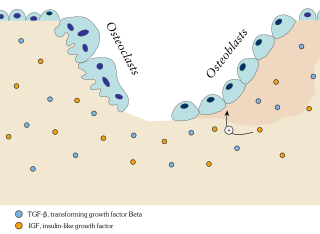
Bone remodeling is a lifelong process where mature bone tissue is removed from the skeleton and new bone tissue is formed. These processes also control the reshaping or replacement of bone following injuries like fractures but also micro-damage, which occurs during normal activity. Remodeling responds also to functional demands of the mechanical loading.

A pathologic fracture is a bone fracture caused by weakness of the bone structure that leads to decrease mechanical resistance to normal mechanical loads. This process is most commonly due to osteoporosis, but may also be due to other pathologies such as: cancer, infection, inherited bone disorders, or a bone cyst. Only a small number of conditions are commonly responsible for pathological fractures, including osteoporosis, osteomalacia, Paget's disease, Osteitis, osteogenesis imperfecta, benign bone tumours and cysts, secondary malignant bone tumours and primary malignant bone tumours.

Comparative foot morphology involves comparing the form of distal limb structures of a variety of terrestrial vertebrates. Understanding the role that the foot plays for each type of organism must take account of the differences in body type, foot shape, arrangement of structures, loading conditions and other variables. However, similarities also exist among the feet of many different terrestrial vertebrates. The paw of the dog, the hoof of the horse, the manus (forefoot) and pes (hindfoot) of the elephant, and the foot of the human all share some common features of structure, organization and function. Their foot structures function as the load-transmission platform which is essential to balance, standing and types of locomotion.
Wolff's law, developed by the German anatomist and surgeon Julius Wolff (1836–1902) in the 19th century, states that bone in a healthy person or animal will adapt to the loads under which it is placed. If loading on a particular bone increases, the bone will remodel itself over time to become stronger to resist that sort of loading. The internal architecture of the trabeculae undergoes adaptive changes, followed by secondary changes to the external cortical portion of the bone, perhaps becoming thicker as a result. The inverse is true as well: if the loading on a bone decreases, the bone will become less dense and weaker due to the lack of the stimulus required for continued remodeling. This reduction in bone density (osteopenia) is known as stress shielding and can occur as a result of a hip replacement. The normal stress on a bone is shielded from that bone by being placed on a prosthetic implant.

In the vertebrate spinal column, each vertebra is an irregular bone with a complex structure composed of bone and some hyaline cartilage, the proportions of which vary according to the segment of the backbone and the species of vertebrate.
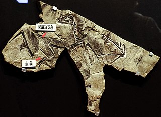
Yi is a genus of scansoriopterygid dinosaurs from the Late Jurassic of China. Its only species, Yi qi, is known from a single fossil specimen of an adult individual found in Middle or Late Jurassic Tiaojishan Formation of Hebei, China, approximately 159 million years ago. It was a small, possibly tree-dwelling (arboreal) animal. Like other scansoriopterygids, Yi possessed an unusual, elongated third finger, that appears to have helped to support a membranous gliding plane made of skin. The planes of Yi qi were also supported by a long, bony strut attached to the wrist. This modified wrist bone and membrane-based plane is unique among all known dinosaurs, and might have resulted in wings similar in appearance to those of bats.
References
- Burr, D.B.; Allen, M.R. (2013). Basic and Applied Bone Biology. Elsevier Science. pp. 23–24. ISBN 978-0-12-391459-0.
| | This human musculoskeletal system article is a stub. You can help Wikipedia by expanding it. |