A reflex, or reflex action, is an involuntary and nearly instantaneous movement in response to a stimulus. A reflex is made possible by neural pathways called reflex arcs which can act on an impulse before that impulse reaches the brain. The reflex is then an automatic response to a stimulus that does not receive or need conscious thought.

The somatic nervous system is the part of the peripheral nervous system associated with the voluntary control of body movements via skeletal muscles.

The cremaster muscle is a muscle that covers the testis and the spermatic cord.

A reflex arc is a neural pathway that controls a reflex. In vertebrates, most sensory neurons do not pass directly into the brain, but synapse in the spinal cord. This allows for faster reflex actions to occur by activating spinal motor neurons without the delay of routing signals through the brain. However, the brain will receive the sensory input while the reflex is being carried out and the analysis of the signal takes place after the reflex action.
The dartos fascia or simply dartos is a layer of connective tissue found in the penile shaft, foreskin, and scrotum. The penile portion is referred to as the superficial fascia of penis or the subcutaneous tissue of penis, while the scrotal part is the dartos proper. In addition to being continuous with itself between the scrotum and the penis, it is also continuous with Colles fascia of the perineum and Scarpa's fascia of the abdomen.

The rectus abdominis muscle, also known as the "abdominal muscle" or "abs", is a paired muscle running vertically on each side of the anterior wall of the human abdomen, as well as that of some other mammals. There are two parallel muscles, separated by a midline band of connective tissue called the linea alba. It extends from the pubic symphysis, pubic crest and pubic tubercle inferiorly, to the xiphoid process and costal cartilages of ribs V to VII superiorly. The proximal attachments are the pubic crest and the pubic symphysis. It attaches distally at the costal cartilages of ribs 5-7 and the xiphoid process of the sternum.

The myometrium is the middle layer of the uterine wall, consisting mainly of uterine smooth muscle cells, but also of supporting stromal and vascular tissue. Its main function is to induce uterine contractions.

The inguinal ligament is a band running from the pubic tubercle to the anterior superior iliac spine. It forms the base of the inguinal canal through which an indirect inguinal hernia may develop.
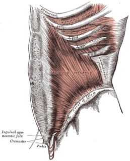
The internal oblique muscle is a muscle in the abdominal wall that lies below the external oblique and just above the transverse abdominal muscles.

The external oblique muscle is the largest and the outermost of the three flat muscles of the lateral anterior abdomen.

The intercostal nerves are part of the somatic nervous system, and arise from the anterior rami of the thoracic spinal nerves from T1 to T11. The intercostal nerves are distributed chiefly to the thoracic pleura and abdominal peritoneum and differ from the anterior rami of the other spinal nerves in that each pursues an independent course without plexus formation.

In anatomy, the abdominal wall represents the boundaries of the abdominal cavity. The abdominal wall is split into the posterior (back), lateral (sides) and anterior (front) walls.
The vacuum exercise is an exercise which involves contracting some internal abdominal muscles, primarily the transverse abdominal muscle, and not as much the diaphragm.
The stretch reflex is a muscle contraction in response to stretching within the muscle. It is a monosynaptic reflex which provides automatic regulation of skeletal muscle length.
Abdominal exercises are those that affect the abdominal muscles.
The cough reflex has both sensory (afferent) mainly via the vagus nerve and motor (efferent) components. Pulmonary irritant receptors in the epithelium of the respiratory tract are sensitive to both mechanical and chemical stimuli. The bronchi and trachea are so sensitive to light touch that slight amounts of foreign matter or other causes of irritation initiate the cough reflex. The larynx and carina are especially sensitive. Terminal bronchioles and even the alveoli are sensitive to chemical stimuli such as sulfur dioxide gas or chlorine gas. Rapidly moving air usually carries with it any foreign matter that is present in the bronchi or trachea. Stimulation of the cough receptors by dust or other foreign particles produces a cough, which is necessary to remove the foreign material from the respiratory tract before it reaches the lungs.
The Golgi tendon reflex is a normal component of the reflex arc of the peripheral nervous system. In a Golgi tendon reflex, skeletal muscle contraction causes the antagonist muscle to simultaneously lengthen and relax. This reflex is also called the inverse myotatic reflex, because it is the inverse of the stretch reflex. Though muscle tension is increasing during the contraction, alpha motor neurons in the spinal cord supplying the muscle are inhibited. However, antagonistic muscles are activated.
An abdominal reflex is a superficial neurological reflex stimulated by stroking of the abdomen around the umbilicus. It can be helpful in determining the level of a CNS lesion. Being a superficial reflex, it is polysynaptic.
