Related Research Articles

The thorax or chest is a part of the anatomy of mammals and other tetrapod animals located between the neck and the abdomen. In insects, crustaceans, and the extinct trilobites, the thorax is one of the three main divisions of the creature's body, each of which is in turn composed of multiple segments.

The mediastinum is the central compartment of the thoracic cavity. Surrounded by loose connective tissue, it is an undelineated region that contains a group of structures within the thorax, namely the heart and its vessels, the esophagus, the trachea, the phrenic and cardiac nerves, the thoracic duct, the thymus and the lymph nodes of the central chest.

The intercostal nerves are part of the somatic nervous system, and arise from the anterior rami of the thoracic spinal nerves from T1 to T11. The intercostal nerves are distributed chiefly to the thoracic pleura and abdominal peritoneum, and differ from the anterior rami of the other spinal nerves in that each pursues an independent course without plexus formation.
Pleuron may refer to:
The arthropod leg is a form of jointed appendage of arthropods, usually used for walking. Many of the terms used for arthropod leg segments are of Latin origin, and may be confused with terms for bones: coxa, trochanter, femur, tibia, tarsus, ischium, metatarsus, carpus, dactylus, patella.

Insect wings are adult outgrowths of the insect exoskeleton that enable insects to fly. They are found on the second and third thoracic segments, and the two pairs are often referred to as the forewings and hindwings, respectively, though a few insects lack hindwings, even rudiments. The wings are strengthened by a number of longitudinal veins, which often have cross-connections that form closed "cells" in the membrane. The patterns resulting from the fusion and cross-connection of the wing veins are often diagnostic for different evolutionary lineages and can be used for identification to the family or even genus level in many orders of insects.

The sternum is the ventral portion of a segment of an arthropod thorax or abdomen.

The mesothorax is the middle of the three segments of the thorax of hexapods, and bears the second pair of legs. Its principal sclerites are the mesonotum (dorsal), the mesosternum (ventral), and the mesopleuron (lateral) on each side. The mesothorax is the segment that bears the forewings in all winged insects, though sometimes these may be reduced or modified, as in beetles (Coleoptera) or Dermaptera, in which they are sclerotized to form the elytra, and the Strepsiptera, in which they are reduced to form halteres that attach to the mesonotum. All adult insects possess legs on the mesothorax. In some groups of insects, the mesonotum is hypertrophied, such as in Diptera, Hymenoptera, and Lepidoptera), in which the anterior portion of the mesonotum forms most of the dorsal surface of the thorax. In these orders, there is also typically a small sclerite attached to the mesonotum that covers the wing base, called the tegula. In one group of insects, the Hemiptera, the dorsal surface of the thorax is typically formed primarily of the prothorax, but also in part by the enlarged posterior portion of the mesonotum, called the scutellum; in the Coleoptera, the scutellum may or may not be visible, usually as a small triangular plate between the elytral bases, thus similar in position to the Hemipteran scutellum. In Diptera and Hymenoptera the mesothoracic scutellum is also distinct, but much smaller than the mesoscutum.

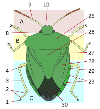
A tergum is the dorsal ('upper') portion of an arthropod segment other than the head. The anterior edge is called the 'base' and posterior edge is called the 'apex' or 'margin'. A given tergum may be divided into hardened plates or sclerites commonly referred to as tergites.

The thoracic wall or chest wall is the boundary of the thoracic cavity.
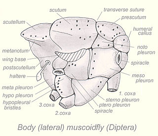
The notopleuron is a region on an insect thorax. Notopleura are useful in characterizing species, particularly, though not uniquely, in the Order Diptera. The notopleuron is a thoracic pleurite situated at the end of the transverse suture of Diptera.
Arthropods are covered with a tough, resilient integument, cuticle or exoskeleton of chitin. Generally the exoskeleton will have thickened areas in which the chitin is reinforced or stiffened by materials such as minerals or hardened proteins. This happens in parts of the body where there is a need for rigidity or elasticity. Typically the mineral crystals, mainly calcium carbonate, are deposited among the chitin and protein molecules in a process called biomineralization. The crystals and fibres interpenetrate and reinforce each other, the minerals supplying the hardness and resistance to compression, while the chitin supplies the tensile strength. Biomineralization occurs mainly in crustaceans. In insects and arachnids, the main reinforcing materials are various proteins hardened by linking the fibres in processes called sclerotisation and the hardened proteins are called sclerotin. The dorsal tergum, ventral sternum, and the lateral pleura form the hardened plates or sclerites of a typical body segment.

Toxomerus marginatus, also known as the margined calligrapher fly, is a common species of hoverfly. It is found in many parts of North America.
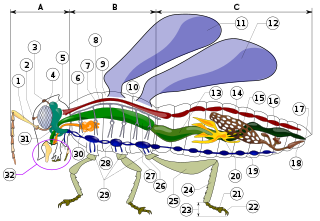
Insect morphology is the study and description of the physical form of insects. The terminology used to describe insects is similar to that used for other arthropods due to their shared evolutionary history. Three physical features separate insects from other arthropods: they have a body divided into three regions, three pairs of legs, and mouthparts located outside of the head capsule. This position of the mouthparts divides them from their closest relatives, the non-insect hexapods, which include Protura, Diplura, and Collembola.
This glossary describes the terms used in formal descriptions of spiders; where applicable these terms are used in describing other arachnids.
The thorax is the midsection (tagma) of the hexapod body. It holds the head, legs, wings and abdomen. It is also called mesosoma or cephalothorax in other arthropods.

Megaphragma is a genus of wasp in the family Trichogrammatidae. It contains some of the smallest known insects, Megaphragma caribea and Megaphragma mymaripenne, which are roughly the same size as some unicellular protozoans.
Most insects reproduce oviparously, i.e. by laying eggs. The eggs are produced by the female in a pair of ovaries. Sperm, produced by the male in one testicle or more commonly two, is transmitted to the female during mating by means of external genitalia. The sperm is stored within the female in one or more spermathecae. At the time of fertilization, the eggs travel along oviducts to be fertilized by the sperm and are then expelled from the body ("laid"), in most cases via an ovipositor.

The pulmonary pleurae are the two flattened sacs ensheathing each lung, locally appearing as two opposing layers of serous membrane separating the lungs from the mediastinum and the inside surfaces of the surrounding chest walls.

Lasioglossum leucozonium, also known as Lasioglossum similis, is a widespread solitary sweat bee found in North America, Europe, Asia, and parts of northern Africa. While now a common bee in North America, population genetic analysis has shown that it is actually an introduced species in this region. This population was most likely founded by a single female bee.
References
- ↑ Zombori, L.; Henrik Steinmann (1999). Dictionary of insect morphology, Volume 4 (Handbuch der Zoologie. Teilband 34). Walter de Gruyter. p. 177. ISBN 978-3-11-014898-5 . Retrieved 2010-04-02.
- ↑ "Pleuron". Merriam-Webster.com Dictionary . Retrieved 16 August 2024.
- ↑ "Episternum". Merriam-Webster.com Dictionary . Retrieved 16 August 2024.
- ↑ "Epimeron". Merriam-Webster.com Dictionary . Retrieved 16 August 2024.
| | This insect anatomy–related article is a stub. You can help Wikipedia by expanding it. |