The maxilla in animals is the upper fixed bone of the jaw formed from the fusion of two maxillary bones. The upper jaw includes the hard palate in the front of the mouth. The two maxillary bones are fused at the intermaxillary suture, forming the anterior nasal spine. This is similar to the mandible, which is also a fusion of two mandibular bones at the mandibular symphysis. The mandible is the movable part of the jaw.
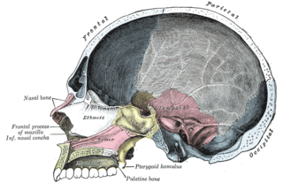
The palatine bones are two irregular bones of the facial skeleton in many animal species. Together with the maxillae they comprise the hard palate.

The inferior nasal concha is one of the three paired nasal conchae in the nose. It extends horizontally along the lateral wall of the nasal cavity and consists of a lamina of spongy bone, curled upon itself like a scroll,. The inferior nasal conchae are considered a pair of facial bones. As the air passes through the turbinates, the air is churned against these mucosa-lined bones in order to receive warmth, moisture and cleansing. Superior to inferior nasal concha are the middle nasal concha and superior nasal concha which arise from the cranial portion of the skull. Hence, these two are considered as a part of the cranial bones.

The vomer is one of the unpaired facial bones of the skull. It is located in the midsagittal line, and articulates with the sphenoid, the ethmoid, the left and right palatine bones, and the left and right maxillary bones. The vomer forms the inferior part of the nasal septum, with the superior part formed by the perpendicular plate of the ethmoid bone. The name is derived from the Latin word for a ploughshare and the shape of the bone.

The internal carotid artery is a major paired artery, one on each side of the head and neck, in human anatomy. They arise from the common carotid arteries where these bifurcate into the internal and external carotid arteries at cervical vertebral level 3 or 4; the internal carotid artery supplies the brain, while the external carotid nourishes other portions of the head, such as face, scalp, skull, and meninges.

The hard palate is a thin horizontal bony plate made up of two bones of the facial skeleton, located in the roof of the mouth. The bones are the palatine process of the maxilla and the horizontal plate of palatine bone. The hard palate spans the alveolar arch formed by the alveolar process that holds the upper teeth.

The middle cerebral artery (MCA) is one of the three major paired arteries that supply blood to the cerebrum. The MCA arises from the internal carotid and continues into the lateral sulcus where it then branches and projects to many parts of the lateral cerebral cortex. It also supplies blood to the anterior temporal lobes and the insular cortices.

The condyloid process or condylar process is the process on the human mandible and some other species' mandibles that ends in a condyle, the mandibular condyle. It is thicker than the coronoid process of the mandible and consists of two portions: the condyle and the constricted portion which supports it, the neck.

In the human mouth, the incisive foramen, also called anterior palatine foramen, or nasopalatine foramen is a funnel-shaped opening in the bone of the oral hard palate immediately behind the incisor teeth where blood vessels and nerves pass. The incisive foramen is continuous with the incisive canal, this foramen or group of foramina is located behind the central incisor teeth in the incisive fossa of the maxilla.

The base of the cartilaginous portion of the auditory tube lies directly under the mucous membrane of the nasal part of the pharynx, where it forms an elevation, the torus tubarius, the torus of the auditory tube, or cushion, behind the pharyngeal orifice of the tube. The torus tubarius is very close to the tubal tonsil, which is sometimes also called the tonsil of (the) torus tubarius. Equating the torus with its tonsil however might be seen as incorrect or imprecise.

The anterior nasal spine, or anterior nasal spine of maxilla, is a bony projection in the skull that serves as a cephalometric landmark. The anterior nasal spine is the projection formed by the fusion of the two maxillary bones at the intermaxillary suture. It is placed at the level of the nostrils, at the uppermost part of the philtrum and rarely fractures.

In human anatomy of the mouth, the palatine process of maxilla, is a thick, horizontal process of the maxilla. It forms the anterior three quarters of the hard palate, the horizontal plate of the palatine bone making up the rest.

The posterior chamber is a narrow space behind the peripheral part of the iris, and in front of the suspensory ligament of the lens and the ciliary processes. The posterior chamber consists of small space directly posterior to the iris but anterior to the lens. The posterior chamber is part of the anterior segment and should not be confused with the vitreous chamber.

The greater palatine nerve is a branch of the pterygopalatine ganglion that carries both general sensory fibres from the maxillary nerve and parasympathetic fibers from the nerve of the pterygoid canal. It descends through the greater palatine canal, emerges upon the hard palate through the greater palatine foramen, and passes forward in a groove in the hard palate, nearly as far as the incisor teeth.

A bronchopulmonary segment is a portion of lung supplied by a specific tertiary bronchus and arteries. These arteries branch from the pulmonary and bronchial arteries, and run together through the center of the segment. Veins and lymphatic vessels drain along the edges of the segment. The segments are separated from each other by layers of connective tissue. Each bronchopulmonary segment is a discrete anatomical and functional unit, and this separation means that a bronchopulmonary segment can be surgically removed without affecting the function of the others.

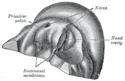
The intermaxillary segment in an embryo is a mass of tissue formed by the merging of tissues in the vicinity of the nose. It is essential for human survival. It is primordial, since in the further development of the embryo this particular mass no longer appears, but parts of it remain in "the intermaxillary portion of the upper jaw, the portion of the upper lip, and the primary palate".

Superior to the anterior portion of the trochlea is a small depression, the coronoid fossa, which receives the coronoid process of the ulna during flexion of the forearm. It is directly adjacent to the radial fossa of the humerus.

The premaxilla is one of a pair of small cranial bones at the very tip of the upper jaw of many animals, usually, but not always, bearing teeth. In humans, they are fused with the maxilla and usually termed as the incisive bone. Other terms used for this structure include premaxillary bone or os premaxillare, and intermaxillary bone or os intermaxillare.

In human anatomy, the mouth is the first portion of the alimentary canal that receives food and produces saliva. The oral mucosa is the mucous membrane epithelium lining the inside of the mouth.
The face and neck development of the human embryo refers to the development of the structures from the third to eighth week that give rise to the future head and neck. They consist of three layers, the ectoderm, mesoderm and endoderm, which form the mesenchyme, neural crest and neural placodes. The paraxial mesoderm forms structures named somites and somitomeres that contribute to the development of the floor of the brain and voluntary muscles of the craniofacial region. The lateral plate mesoderm consists of the laryngeal cartilages. The three tissue layers give rise to the pharyngeal apparatus, formed by six pairs of pharyngeal arches, a set of pharyngeal pouches and pharyngeal grooves, which are the most typical feature in development of the head and neck. The formation of each region of the face and neck is due to the migration of the neural crest cells which come from the ectoderm. These cells determine the future structure to develop in each pharyngeal arch. Eventually, they also form the neurectoderm, which forms the forebrain, midbrain and hindbrain, cartilage, bone, dentin, tendon, dermis, pia mater and arachnoid mater, sensory neurons, and glandular stroma.